
Learn about methods used to map the secretome; secretome mapping; its methods; multiplex assays; proteomics; bioinformatics tools; and applications in cancer research, biomarkers, and precision medicine.
Introduction
Imagine your neighborhood has a group of people who quietly leave useful items outside their homes every morning—fresh milk, newspapers, or food parcels. By looking at what is left outside, you can understand who is contributing and what they are doing, even without entering their houses.
Scientists study the secretome in a similar way. Instead of looking inside cells, they analyze the proteins, peptides, and other molecules that cells release into their surroundings. Mapping the secretome helps researchers identify these secreted molecules, understand how cells communicate, and discover biomarkers for diseases and potential therapeutic targets.
Methods used to map the secretome are techniques that identify, measure, and analyze all the molecules secreted by cells into their environment.
Secretome analysis is widely used to study different cell types, including hematopoietic stem cells, to understand how they communicate and support tissue regeneration.
Methods used to map the secretome
Scientists can evaluate the intricate alterations in gene and protein expression brought on by stimuli like antigen or cytokine activation by using the linked fields of proteomics and genomics. It is now possible to evaluate and show vast amounts of data about the expression of specific proteins as well as the derivation and readout of genes in various cells and creatures in ways that were not possible for scientists only a few years ago.
The mapping of proteins released by different cell types has recently been added to the field of proteomics. A cell’s secretome is the collection of proteins it secretes; more officially, it is defined as “proteins released by a cell, tissue, or organism through classical and nonclassical secretion mechanisms.”
The idea of using the secretome to diagnose and detect different forms of cancer initially piqued the curiosity of scientists. They reasoned that the proteins produced in the serum or other tissue fluids could serve as a biological marker for particular types of tumors. If specific proteins can be demonstrated to be secreted at high concentrations only under malignant situations, then quick and low-cost assays may be created that could screen for tumors early on, when they are still treatable.
Given the variety of mutations linked to the development of cancer, it is surprisingly challenging to create such tumor-specific profiles of secreted proteins. However, the ability to diagnose a tumor at an early stage using only a serum sample provides intense motivation, and many such attempts are ongoing.
Since then, the methods employed to identify a set of cancer secretomes have been applied to research on numerous other non-malignant cell populations for which a secretome description would be a valuable analytical tool. These populations include fat cells, immune system cells, and stem cells. Cytokine biology is an excellent target for such a global strategy because of the variety of cytokines that can be released by a single cell and the way that cytokine activities can interact at the level of the target cell.
The subject of how the human cytomegalovirus stimulates the development of new blood vessels (angiogenesis) was addressed in a recent secretome investigation (Botto et al., 2011). Angiogenesis was discovered to be induced by virus-free supernatant from virus-infected endothelial cells. Several cytokines, including IL-8, GM-CSF, and IL-6, were found in the secretome analysis of the infected endothelial cell supernatant. It was subsequently demonstrated that the addition of a blocking anti-IL-6 antibody concurrently with the virus-free supernatant inhibited its angiogenic activity, indicating that the IL-6 activity in the supernatant was the primary cause of the new blood vessel formation.
The requirement for many cells to develop in a tissue culture fluid supplemented with serum, which is itself a complex mixture of proteins, is one challenge that is commonly encountered when attempting to investigate the secretome of a certain kind of cell. In this instance, it’s critical to differentiate between the proteins that were initially in the serum and those that were released by the cells being studied. There are a number of methods to distinguish between secreted proteins and those from the tissue culture media, such as introducing secretion inhibitors to some cultures and then comparing the pro-teins found in the culture supernatant with and without the inhibitors.
An alternative method for separating freshly generated proteins from preexisting proteins in the growth medium is to cultivate the cells in the presence of radioisotopes, such as 35S methionine, that exclusively label newly synthesized proteins.
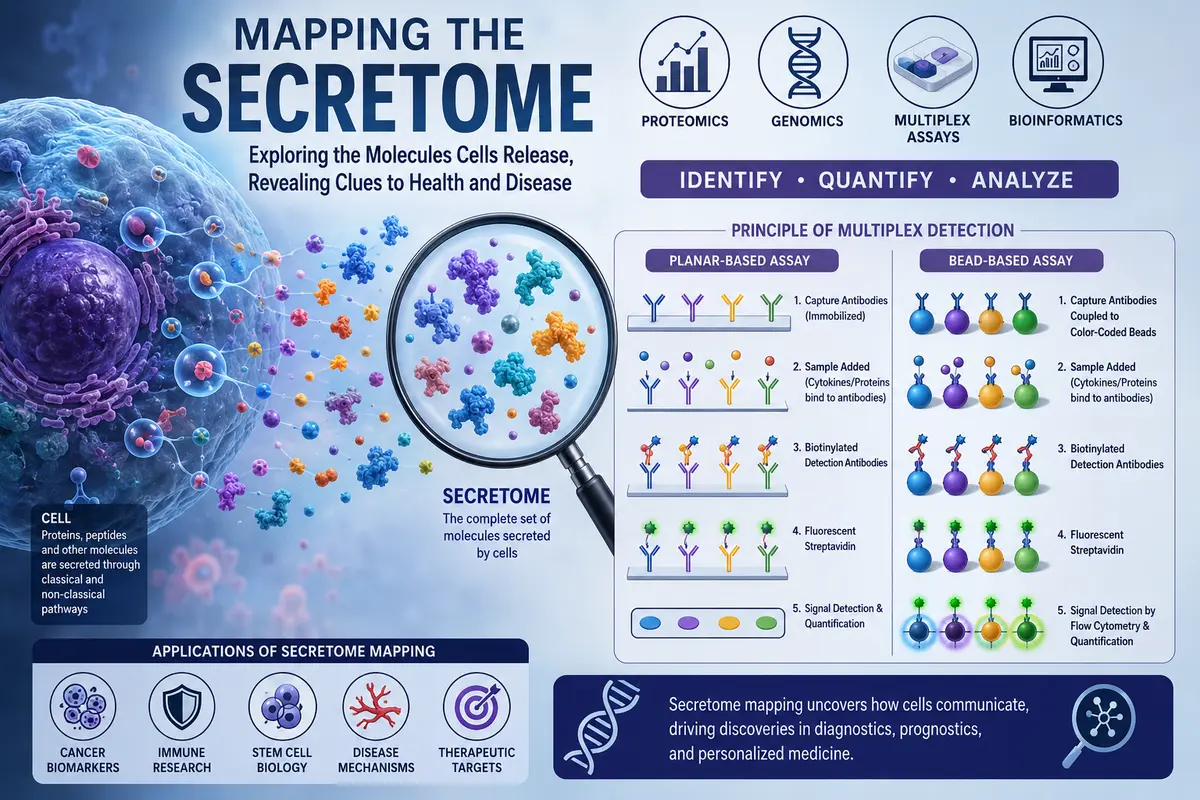
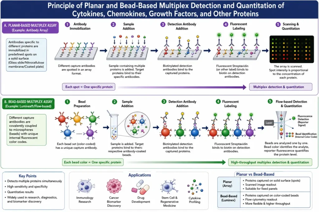
Two different kinds of multiplex measurements may be employed in situations like the one mentioned above, when a cell line is being examined to see if it secretes a collection of cytokines for which antibody assays already exist. Both of these methods make use of antibodies to the range of cytokines that need to be examined, affixed to a solid-phase support of some kind. Glass, a membrane, or a collection of beads with each antibody affixed to a different-colored bead could serve as this support. After adding the tissue culture fluid sample to the solid phase antibody and washing away any extra fluid, biotinylated antibodies are added.
In experiments like these, biotin, a tiny molecule, is utilized to couple two molecules together because of its exceptionally high affinity for the protein streptavidin. Following antibody binding, surplus biotin-nylated antibodies are eliminated by washing, and fluorescent streptavidin is added to measure cytokine concentrations by binding to the biotin. The presence of the cytokine in the sample is shown by a fluorescent signal, and the signal’s intensity represents its concentration. The fluorescence linked to each cytokine can be identified since each bead fluoresces at a distinct wavelength.
Numerous bioinformatics tools that are specifically useful for secretome analysis have been created. Among these is SignalP, which locates signal peptide cleavage sites in bacterial and eukaryotic proteins and detects the presence of signal peptides. Furthermore, nonclassically secreted proteins can be predicted using SecretomeP.
The protein sequence is used by a number of bioinformatics tools, such as TargetP and Protein Prowler, to estimate the subcellular location of the protein. Lastly, the Ingenuity Pathway. Analysis enables the researcher to forecast the protein of interest’s involvement in functional networks and to look for protein interaction partners.
Principle of planar and bead-based multiplex detection and quantitation of cytokines, chemokines, growth factors, and other proteins.
Conclusion
Secretome mapping is a powerful approach for identifying and analyzing the proteins released by cells. Using techniques such as proteomics, multiplex immunoassays, radioisotope labeling, and bioinformatics tools, researchers can better understand cell-to-cell communication, disease mechanisms, and immune responses. Secretome analysis has become especially valuable in cancer research, stem cell biology, immunology, and biomarker discovery, enabling earlier disease detection and the identification of new therapeutic targets.
As analytical technologies continue to improve, secretome mapping is expected to play an increasingly important role in precision medicine, diagnostics, and personalized treatment strategies.
Many cytokines detected during secretome analysis are produced by different components of the immune system.
FAQs
1. What is a secretome?
Answer: A secretome is the complete collection of proteins, peptides, cytokines, growth factors, and other molecules that are secreted by a cell, tissue, or organism into its surrounding environment through classical and non-classical secretion pathways.
2. What is secretome therapy?
Answer: Secretome therapy is a regenerative treatment that uses the bioactive molecules (such as growth factors, cytokines, exosomes, and proteins) secreted by stem cells to promote tissue repair, reduce inflammation, and support healing without transplanting the stem cells themselves.
3. Secretome vs. exosome: Which is better?
Answer: Neither is universally better—it depends on the application.
The secretome contains all molecules released by cells, including growth factors, cytokines, proteins, and exosomes, making it suitable for broad tissue repair and regeneration.
Exosomes are tiny extracellular vesicles within the secretome that deliver specific proteins, lipids, and RNA to target cells, offering more targeted cell-to-cell communication.
In general, secretome is preferred for comprehensive regenerative effects, while exosomes may be better for targeted therapeutic applications and precision medicine.
4. What is a secretome stem cell?
Answer: Stem cell secretome is the collection of bioactive molecules—including growth factors, cytokines, proteins, peptides, extracellular vesicles, and exosomes—released by stem cells. These molecules help promote tissue repair, reduce inflammation, stimulate regeneration, and support cell-to-cell communication without using the stem cells themselves.
5. What is the secretome of a bacterium?
Answer: The bacterial secretome is the complete set of proteins, enzymes, toxins, and other molecules secreted by a bacterium into its surrounding environment. These secreted molecules help bacteria obtain nutrients, communicate, interact with host cells, and cause infection or disease.
References
- Owen, J. A., Punt, J., Stranford, S. A., & Jones, P. P. (2013). Kuby Immunology (7th ed.). W. H. Freeman and Company.
- Agrawal, G. K., & Rakwal, R. (2011). Rice Proteomics: A Cornerstone for Cereal Food Crop Proteomes. John Wiley & Sons.
- Botto, S., et al. (2011). Human cytomegalovirus infection induces a secretome that promotes angiogenesis through interleukin-6. Proceedings of the National Academy of Sciences, 108(31), 13030–13035.
- Greenbaum, D., Luscombe, N. M., Jansen, R., Qian, J., & Gerstein, M. (2001). Interrelating different types of genomic data, from proteome to secretome: ‘Omics’ and beyond. Genome Research, 11(9), 1463–1468.
- Hathout, Y. (2007). Approaches to the study of the cell secretome. Expert Review of Proteomics, 4(2), 239–248.
- Tjalsma, H., Bolhuis, A., Jongbloed, J. D. H., Bron, S., & van Dijl, J. M. (2000). Signal peptide-dependent protein transport in Bacillus subtilis: A genome-based survey of the secretome. Microbiology and Molecular Biology Reviews, 64(3), 515–547.
- Bendtsen, J. D., Jensen, L. J., Blom, N., von Heijne, G., & Brunak, S. (2004). Feature-based prediction of non-classical and leaderless protein secretion. Protein Engineering, Design & Selection, 17(4), 349–356.
- Almagro Armenteros, J. J., et al. (2019). SignalP 5.0 improves signal peptide predictions using deep neural networks. Nature Biotechnology, 37(4), 420–423.
- Emanuelsson, O., Nielsen, H., Brunak, S., & von Heijne, G. (2000). Predicting subcellular localization of proteins based on their N-terminal amino acid sequence. Journal of Molecular Biology, 300(4), 1005–1016.
- Diks, S. H., et al. (2004). The prospects of secretome analysis in biomarker discovery. Journal of Cellular Biochemistry, 93(4), 665–674.