
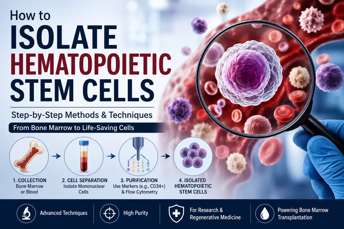
Learn how to isolate hematopoietic stem cells using simple, step-by-step methods. Understand bone marrow collection, cell separation, and purification techniques.
Introduction
Imagine you have a huge basket filled with different kinds of fruits—apples, oranges, bananas, grapes, and mangoes—but your teacher asks you to pick only the apples. You wouldn’t throw away the whole basket. Instead, you would carefully identify the apples based on their color, size, and shape and separate them from the rest.
Scientists do something very similar when they isolate hematopoietic stem cells (HSCs). The bone marrow and blood contain millions of different types of cells, but only a tiny fraction are hematopoietic stem cells. These special cells are responsible for producing all the different types of blood cells throughout a person’s life. To study them or use them for medical treatments, scientists must first identify and separate these rare cells from the large mixture.
Think of it like finding a few VIP guests in a crowded stadium. The VIPs are present, but they need to be identified using special passes. Similarly, hematopoietic stem cells have unique surface markers (such as CD34 in humans) that act like identification badges. Scientists use these markers and advanced laboratory techniques to isolate HSCs accurately.
In this topic, we will learn the different methods used to isolate hematopoietic stem cells, why this process is important, and how these cells are used in research, regenerative medicine, and bone marrow transplantation.
Isolating Hematopoietic Stem Cells
By the 1960s researchers knew that hematopoietic stem cells (HSCs) existed and were a rare population in the bone marrow. But they had no idea what made them different from all the other millions of cells that packed the bone marrow. It was obvious that to truly understand how these amazing cells generate all other blood cells and to take advantage of this potential for clinical use, investigators would have to find a way to isolate HSCs.
But how does one find a thing so rare, where the only difference is its function, its ability to produce all blood cells? Investigators developed a variety of strategies that changed rapidly with each advance in technology, particularly the advent of monoclonal antibodies and flow cytometry.
Whatever way you go about trying to isolate a cell, it is very important to have a reliable experimental assay that can tell you that the cells that you have teased out are indeed the ones you are looking for. To prove that you have enriched or purified an HSC, you have to show, basically, that it can proliferate and give rise to all blood cell types in an animal over the long term. Most of the original assays developed to demonstrate this are still in use.
They include colony formation assays, where the ability of individual cells to proliferate (and differentiate) is assayed by looking for evidence of cell division either in vitro (on plates) or in vivo (in the spleens of irradiated mice). But the strongest evidence of successful isolation of HSCs is that they can restore the blood cells and immune system of a lethally irradiated animal and thus save its life. This can be done for mouse stem cells by injecting stem cell candidates into irradiated mice and seeing if they can give survival and repopulate all blood cell types.
The development of an SCID-hu (man) mouse model that accepts human hematopoietic stem cells has greatly enhanced the ability of investigators to demonstrate the pluripotentiality of candidate human stem cell populations.
In the 1970s, investigators were not able to easily compare differences in protein and gene expression among single cells, so they had to try to distinguish cell types based on other physical and structural features. It was only with the arrival of monoclonal antibodies into research repertoires that serious consideration could be given to the purification of a stem cell. Monoclonal antibodies can be produced against virtually any protein, lipid, or carbohydrate. Monoclonal antibodies can be covalently tagged with gold particles, enzymes, or fluorochromes and visualized by microscopy to show where they bind.
Researchers believed that HSCs were unlikely to express mature blood cell-specific proteins in the early 1980s. They trapped and extracted them from bone marrow cell suspensions using monoclonal antibodies produced against several mature cells. They did this by first using a technique known as panning, in which the heterogeneous pool of cells was incubated with antibodies attached to a plastic, and the cells that did not adhere were then dislodged and poured off.
In fact, the cells with HSC potential that did not adhere to the antibodies were enriched (in some cases by several thousandfold). This form of negative selection against mature cell lineages is still effective and is now called “lineage” or “Lin” selection; cells that are enriched using this technique are called “Lin.”
In order to positively select hematopoietic stem cells among the many bone marrow cell types, researchers also sought to find surface chemicals unique to these cells. A monoclonal antibody produced against a tumor of primitive blood cell types (acute myeloid leukemia) was used to identify the first protein that recognized human HSCs, now known as CD34.
The flow cytometer offers the most effective way to extract a rare population from a diverse group of cells, even though one can positively select cells from a diverse population using the panning techniques mentioned above (or by using its more modern variant where cells are applied to columns of resin-bound monoclonal antibodies or equivalents). Immunology and clinical medicine have been transformed by this device, which was created by the Herzenberg laboratory and its multidisciplinary team of researchers and innovators. To put it briefly, it is a device that enables the identification, separation, and recovery of individual cells from a variety of cell pools based on the profiles of proteins and/or genes they express.
To put it briefly, cells from a heterogeneous solution are “stained with” monoclonal antibodies (or other compounds) that couple to different fluorochromes and attach to different characteristics. The intensities of the various wavelengths that each individual cell emits are recorded when these cells flow in single file in front of lasers that activate the various fluorochromes. It is possible to physically separate cells that exhibit desired levels of expression of particular antigens (such as CD34) from other cells and recover them for additional research.
Irv Weissman and his labs found in the late 1980s that mouse hematopoietic stem cells could be distinguished from more mature cells by variations in the production of the T-cell marker Thy protein and, subsequently, Sca pro-tein. His laboratory developed one of the most effective methods for hematopoietic stem cell enrichment by combining both positive and negative selection strategies. The method was refined as additional surface molecules were discovered. Nowadays, the Lin Sca-1+ c-Kit+ (“LSK”) phenotype is the most used way to identify HSCs. Even this subgroup, which makes up less than 1% of bone marrow cells, has been shown to exhibit phenotypic and functional heterogeneity.
SLAM proteins and other surface indicators that can differentiate between these subpopulations are still being identified. In the end, we will be able to clearly identify, isolate, and manipulate what remains the holy grail of HSC research—the long-term stem cell that can both self-renew and give rise to all blood cell types—thanks to the continued synergy between technical advancements and experimental strategies.
Classic Experiment
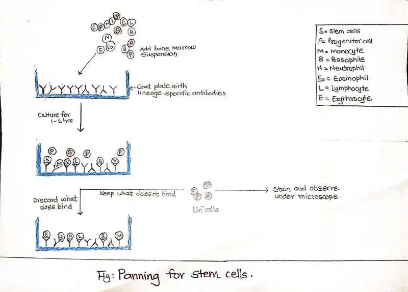
Antibodies produced against adult blood cells and a procedure known as panning, in which cells are cultured in plastic plates coated with antibodies, were used in early methods to isolate HSCs. In particular, a solution of bone marrow was placed onto plastic plates coated with antibodies capable of binding certain mature (“lineage positive”) blood cells. The immature, “lineage-negative” blood cells that did not adhere were removed from the plate after a wait of one to two hours.
The majority of mature blood cells adhered securely to the plate. Hematopoietic stem cells were more prevalent in the cells that did not adhere. The first picture of human bone marrow cells enriched for hematopoietic stem cells through panning was produced as a result of this procedure. S stands for stem cell, P for progenitor cell, M for monocyte, B for basophil, N for neutrophil, Eo for eosinophil, L for lymphocyte, and E for erythrocyte.
Current approaches for enrichment of the pluripotent stem cells from bone marrow.
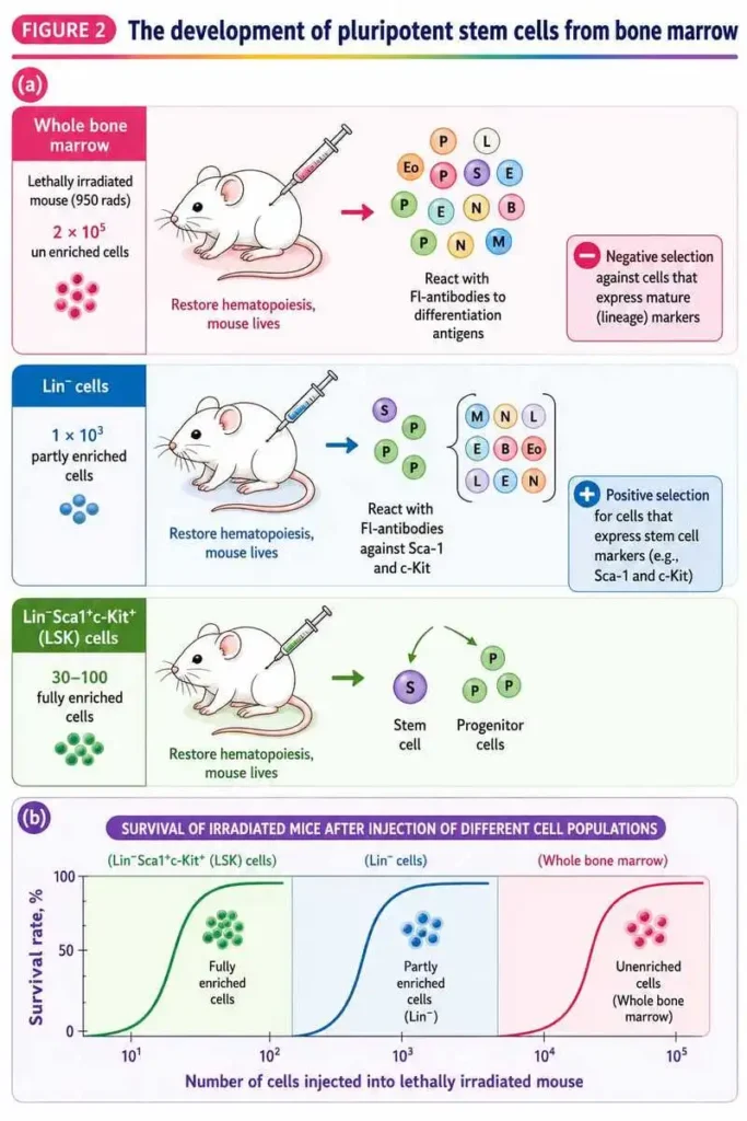
current methods for bone marrow-derived pluripotent stem cell enrichment. Irv Weissman and associates created a schematic of the kind of stem cell enrichment that is currently widely used. (a) After treatment with fluorescently labeled antibodies (Fl-antibodies) specific for membrane molecules expressed on differentiated (mature) lineages but absent from undifferentiated stem cells (S) and progenitor cells (P), differentiated hematopoietic cells (white) are removed (negative selection) from whole bone marrow.
This is followed by (2) retention (positive selection) of cells within the resulting partially enriched preparation that bound to antibodies specific for two early differentiation antigens, Sca-1 and c-Kit. “Lineage-minus,” or Lin, populations are cells that have been enhanced by the elimination of differentiated cells.
LSK (for Lin Sca-1 c-Kit) cells are those that have been further enhanced by positive selection based on Sca-1 and c-Kit expression. M stands for monocyte, B for basophil, N for neutrophil, Eo for eosinophil, L for lymphocyte, and E for erythrocyte. (b) The capacity of stem cell preparations to repair hematopoiesis in mice exposed to fatal radiation is used to gauge their enrichment. The only creatures that survive are those that undergo hematopoiesis. The number of injected cells required to restore hematopoiesis decreases as stem cell enrichment progresses (from total bone marrow to Lin populations to LSK populations). This process can result in a total enrichment of roughly 1000 times.
Conclusion
The isolation of hematopoietic stem cells (HSCs) is one of the most significant achievements in stem cell biology and modern medicine. Since HSCs are extremely rare in bone marrow and blood, scientists rely on advanced techniques such as negative selection (Lin⁻ selection), positive selection using CD34 and other surface markers, panning, magnetic cell separation, and flow cytometry (FACS) to identify and purify these cells accurately.
Over the years, technological advancements and the discovery of specific cell surface markers like CD34, Sca-1, c-Kit, and SLAM proteins have greatly improved the efficiency of HSC isolation. These methods allow researchers to obtain highly purified stem cell populations for laboratory studies and clinical applications.
Today, isolated hematopoietic stem cells play a vital role in stem cell transplantation, bone marrow transplantation, treatment of blood disorders, regenerative medicine, gene therapy, cancer research, and immunology. As research continues, newer technologies are expected to provide even more precise, faster, and safer methods for isolating long-term hematopoietic stem cells, opening new possibilities for personalized medicine and life-saving therapies.
In summary, the ability to isolate hematopoietic stem cells is the foundation of stem cell research and blood-related therapies, enabling scientists and clinicians to better understand, repair, and regenerate the human blood and immune system.
For the latest updates in stem cell science, visit the International Society for Stem Cell Research (ISSCR).
FAQs
1. Where are hematopoietic stem cells located?
Answer: Hematopoietic stem cells (HSCs) are primarily located in the bone marrow, especially in the pelvis, sternum (breastbone), ribs, and vertebrae of adults. They are also found in peripheral blood (in small numbers), umbilical cord blood, and the fetal liver during embryonic development.
2. What is the function of hematopoietic stem cells?
Answer: Hematopoietic stem cells (HSCs) are responsible for producing and replenishing all types of blood cells throughout life. They have the unique ability to self-renew (make more stem cells) and differentiate into red blood cells, white blood cells, and platelets, helping maintain normal blood formation, oxygen transport, immunity, and blood clotting.
3. What cells are used for hematopoietic stem cell transplantation?
Answer: Hematopoietic stem cell transplantation (HSCT) uses hematopoietic stem cells (HSCs), which are immature blood-forming cells capable of producing all types of blood cells. These stem cells are collected from three main sources:
Bone marrow
Peripheral blood (mobilized stem cells)
Umbilical cord blood
These HSCs are transplanted into patients to restore healthy blood cell production after diseases such as leukemia, lymphoma, aplastic anemia, and certain genetic blood disorders.
4. What are hematopoietic stem cells?
Answer: Hematopoietic stem cells (HSCs) are multipotent stem cells that give rise to all types of blood cells, including red blood cells, white blood cells, and platelets. They have two unique abilities: self-renewal, which allows them to produce more stem cells, and differentiation, which enables them to develop into specialized blood cells. HSCs are primarily found in the bone marrow and play a vital role in maintaining a healthy blood and immune system throughout life.
5. Where are hematopoietic stem cells found?
Answer: Hematopoietic stem cells are mainly found in the bone marrow, but they are also present in peripheral blood, umbilical cord blood, and the fetal liver during development.
References
Books
- Alberts, B., Johnson, A., Lewis, J., Morgan, D., Raff, M., Roberts, K., & Walter, P. (2022). Molecular Biology of the Cell (7th ed.). W. W. Norton & Company.
- Kumar, V., Abbas, A. K., & Aster, J. C. (2020). Robbins & Cotran Pathologic Basis of Disease (10th ed.). Elsevier.
- Murphy, K., & Weaver, C. (2022). Janeway’s Immunobiology (10th ed.). Garland Science.
- Lodish, H., Berk, A., Kaiser, C. A., et al. (2021). Molecular Cell Biology (9th ed.). W. H. Freeman.
- Cooper, G. M., & Hausman, R. E. (2019). The Cell: A Molecular Approach (8th ed.). Oxford University Press.
Research Article
- Weissman, I. L. (2000). Stem cells: Units of development, units of regeneration, and units in evolution. Cell, 100(1), 157–168. https://doi.org/10.1016/S0092-8674(00)81692-X
- Orkin, S. H., & Zon, L. I. (2008). Hematopoiesis: An evolving paradigm for stem cell biology. Cell, 132(4), 631–644. https://doi.org/10.1016/j.cell.2008.01.025
- Morrison, S. J., & Spradling, A. C. (2008). Stem cells and niches: Mechanisms that promote stem cell maintenance throughout life. Cell, 132(4), 598–611. https://doi.org/10.1016/j.cell.2008.01.038
- Seita, J., & Weissman, I. L. (2010). Hematopoietic stem cell: Self-renewal versus differentiation. Wiley Interdisciplinary Reviews: Systems Biology and Medicine, 2(6), 640–653. https://doi.org/10.1002/wsbm.86
- Doulatov, S., Notta, F., Laurenti, E., & Dick, J. E. (2012). Hematopoiesis: A human perspective. Cell Stem Cell, 10(2), 120–136. https://doi.org/10.1016/j.stem.2012.01.006