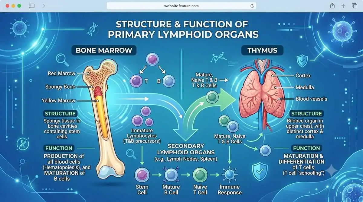
Learn what the structure and function of primary lymphoid organs are, including the bone marrow and thymus. Understand how these organs produce and mature B and T lymphocytes for a strong immune system.
Introduction: What Is the Structure and Function of Primary Lymphoid Organs?
Our body is constantly exposed to germs like bacteria and viruses every day. Just as a school trains students before they start their careers, the body also has special organs that train immune cells to fight infections. These organs are called primary lymphoid organs.
The bone marrow produces blood cells and matures B cells, while the thymus helps T cells mature and learn how to protect the body. Together, these organs prepare the immune system to recognise and destroy harmful germs.
Imagine a police training academy. New recruits first receive training before they are sent out to protect the public. Similarly, bone marrow and the thymus act as training centres, where immune cells are produced and trained before they are released into the body to fight diseases.
For a detailed overview of the immune system and lymphoid organs, learn more about the human immune system from NCBI Bookshelf.
Primary Lymphoid Organs
The structural arrangement and cellular activity of specific anatomic microenvironments known as stem cell niches determine any stem cell’s capacity for self-renewal and differentiation.
Usually, a supporting network of stromal cells populates these isolated areas. Soluble and membrane-bound proteins that control cell survival, proliferation, differentiation, and trafficking are expressed by stem cell niche stromal cells.
Throughout the course of embryonic development, the organs with microenvironments that promote haematopoietic stem cell differentiation actually undergo changes.
But during the middle to late stages of pregnancy, HSCs settle in the bone marrow, which continues to be the principal location for haematopoiesis throughout adulthood. All erythroid and myeloid cells, as well as B lymphocytes in humans and mice, mature in the bone marrow.
HSCs can spontaneously recirculate between the bone marrow and other tissues and are also present in blood. The procedure for transplanting blood cell progenitors from donors into patients who are deficient (such as those who have received chemotherapy) has been made simpler by this observation.
In the past, it was usually necessary to aspirate bone marrow from the donor—a painful procedure requiring anaesthesia—but now it is occasionally feasible to use enriched haematopoietic precursors from donor blood, which is much easier to get.
T lymphocytes do not finish developing in the bone marrow, in contrast to B lymphocytes. To become functional cells, T lymphocyte precursors must leave the bone marrow and move to the special microenvironments offered by the thymus, the other key lymphoid organ.
Primary lymphoid organs are the organs where lymphocytes (B cells and T cells) are produced and mature. In humans, there are two primary lymphoid organs:
- Bone Marrow
- Thymus
1. Bone Marrow
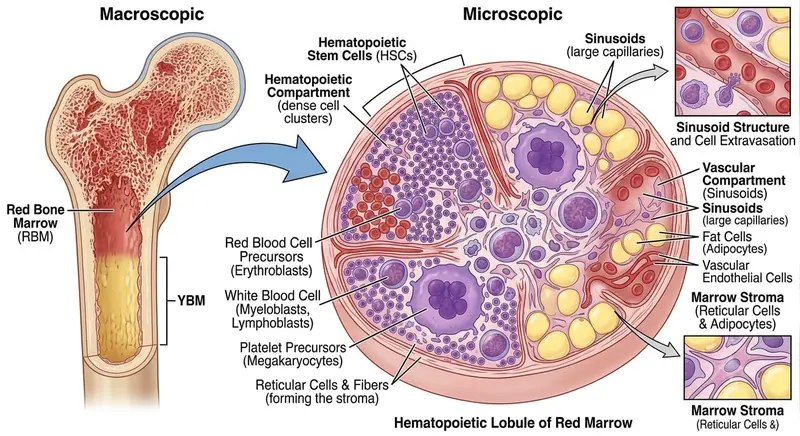
Bone marrow is a soft, highly vascular connective tissue found inside the cavities of bones. It is the primary site of haematopoiesis (blood cell formation) and contains stem cells that produce red blood cells, white blood cells, and platelets.
Structure of Bone Marrow
i) Compact Bone
- Hard outer layer of the bone.
- Provides strength and protection.
ii) Spongy (Cancellous) Bone
- Located beneath the compact bone.
- Contains numerous trabeculae with spaces filled by red bone marrow.
iii) Bone Marrow Cavity (Medullary Cavity)
- Central cavity of long bones.
- Contains yellow bone marrow in adults.
- In children, it is mostly filled with red bone marrow.
iv) Red Bone Marrow
- Found in: flat bones (sternum, ribs, pelvis, skull); vertebrae ends (epiphyses) of long bones; rich in haematopoietic stem cells (HSCs).
- Produces: Red blood cells (RBCs), white blood cells (WBCs), platelets
v) Yellow Bone Marrow
- Mainly composed of adipose (fat) cells.
- Acts as an energy reserve.
- Can convert back into red marrow during severe blood loss or anaemia.
vi) Hematopoietic Stem Cells (HSCs)
- Multipotent stem cells present in red marrow.
- Differentiate into all blood cell types.
vii) Bone Marrow Sinusoids
- Wide, thin-walled blood vessels.
- Allow newly formed blood cells to enter the bloodstream.
viii) Stromal Cells
- Supportive cells that form the bone marrow microenvironment: Reticular cells, fibroblasts, macrophages, endothelial cells, adipocytes (fat cells)
- They provide structural support and release growth factors necessary for blood cell development.
Working of Bone Marrow
Haematopoietic stem cells can self-renew and differentiate into myeloid cells and B lymphocytes thanks to niches provided by the bone marrow.
Haematopoietic stem cells (HSCs) self-renew and differentiate into adult blood cells in the bone marrow, a main lymphoid organ. The long bones (femur and humerus), hip bones (ileum), and sternum are typically the most active sites of haematopoiesis, despite the fact that all bones contain marrow.
In addition to producing and renewing blood cells, the bone marrow is in charge of preserving the pool of HSCs for the duration of an adult vertebrate’s life.
Several cell types that coordinate HSC development are found in the adult bone marrow, the paradigmatic adult stem cell niche.
These include (1) osteoblasts, which are versatile cells that both produce bone and regulate HSC differentiation; (2) endothelial cells, which line blood vessels and also regulate HSC differentiation; (3) reticular cells, which send processes connecting cells to bone and blood vessels; and, surprisingly, (4) sympathetic neurones, which can regulate the release of haematopoietic cells from the bone marrow.
The bone marrow is densely packed with haematopoietic and stromal cells at every stage of differentiation, as shown in a microscopic cross-section. However, the efficiency of haematopoiesis declines with ageing as fat cells progressively replace 50% or more of the bone marrow compartment.
An HSC’s decisions are mostly influenced by the cues it receives from its surroundings. Although the bone marrow is full with haematopoietic cells at every stage of development, each myeloid and lymphoid subtype’s progenitors most likely mature in unique environmental micro-niches inside the bone marrow.
We are continually learning about the microenvironments in the bone marrow that sustain particular stages of haematopoiesis.
However, there is evidence that the vascular niche—the area directly surrounding blood arteries and in contact with endothelial cells—and the endosteal niche—the area directly surrounding the bone and in contact with bone-producing osteoblasts—play distinct functions.
Quiescent HSCs appear to occupy the endosteal niche in close proximity to osteoblasts, which control the proliferation of stem cells. HSCs that have been mobilised to leave the endosteal niche to either differentiate or circulate appear to occupy the vascular niche.
Furthermore, a cell appears to migrate closer to the more central areas of the bone and farther away from its supporting osteoblasts as it becomes more differentiated.
For instance, the most mature B cells have relocated into the more central sinuses of the bone marrow, which are well-served by blood arteries, whereas the most immature B cells are located closest to the endosteum and osteoblasts.
Lastly, it’s critical to understand that the bone marrow serves as a place for both the creation of lymphoid and myeloid cells as well as the return of fully developed lymphoid and myeloid cells. Plasma cells, which are mature B cells that secrete antibodies, may potentially settle down permanently in the bone marrow.
Therefore, whole bone marrow transplants contain mature, functional cells in addition to stem cells, which can both benefit and harm the transplant process.
Function of Bone Marrow
Bone marrow is a soft, spongy tissue found inside bones. It is the primary haematopoietic (blood-forming) organ and plays a vital role in blood cell production, immunity, and fat storage. Supportive cells that form the bone marrow microenvironment:
i) Haematopoiesis (Blood Cell Formation)—The primary function of bone marrow is the production of blood cells from haematopoietic stem cells (HSCs).
It produces red blood cells (RBCs), which transport oxygen. White Blood Cells (WBCs), which protect the body against infections. Platelets (thrombocytes), which help in blood clotting.
ii) Production of Immune Cells—Bone marrow produces B lymphocytes (B cells), which mature in the bone marrow. It also produces T-cell precursors, which migrate to the thymus for maturation. Produces other immune cells such as neutrophils, monocytes, eosinophils, and basophils.
iii) Storage of Fat—Yellow bone marrow stores fat (adipose tissue). Serves as an energy reserve during periods of starvation or illness.
iv) Stem Cell Reservoir—Bone marrow contains haematopoietic stem cells, which continuously replace old and damaged blood cells throughout life.
v) Release of Mature Blood Cells—Mature blood cells pass through bone marrow sinusoids into the bloodstream to circulate throughout the body.
vi) Removal of Old or Abnormal Cells—Macrophages present in the bone marrow remove damaged, dead, or defective blood cells, helping maintain healthy blood composition.
vii) Role in Bone Marrow Transplantation—Bone marrow is a rich source of stem cells used in bone marrow (stem cell) transplantation to treat diseases such as leukaemia, lymphoma, aplastic anaemia, and some inherited blood disorders.
2. Thymus
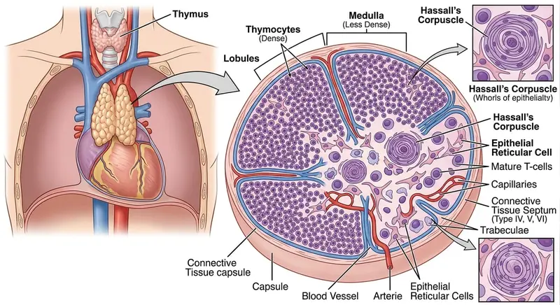
One of the main lymphoid organs where T cells develop is the thymus. The thymus is a primary lymphoid organ responsible for the development, maturation, and selection of T lymphocytes (T cells).
It is located in the upper anterior mediastinum, behind the sternum and above the heart. The thymus is largest during childhood and gradually shrinks after puberty, a process known as thymic involution.
Structure of Thymus
i) Capsule
- The thymus is enclosed by a thin connective tissue capsule.
- The capsule sends inward projections called trabeculae (septa) that divide the gland into many lobules.
ii). Lobules—Each thymic lobule has two distinct regions: a. Cortex (Outer Dark Region), b. Medulla (Inner Light Region)
a. Cortex (Outer Dark Region)
- Dark-staining due to numerous immature T lymphocytes (thymocytes).
- It contains immature T cells, cortical thymic epithelial cells (cTECs), and macrophages.
- Function: T-cell proliferation, formation of T-cell receptors (TCRs), positive selection of functional T cells.
b. Medulla (Inner Light Region)
- Lighter staining because it contains fewer lymphocytes.
- It contains mature T cells, medullary thymic epithelial cells (mTECs), dendritic cells, macrophages, and Hassall’s corpuscles.
- Function: Negative selection to eliminate self-reactive T cells, final maturation of T cells.
iii). Hassall’s Corpuscles
- Unique structures found only in the medulla.
- Made of concentric layers of epithelial cells.
- Help in the maturation of dendritic cells and the development of immune tolerance.
iv) Blood-Thymus Barrier
- Present mainly in the cortex.
- Prevents immature T cells from exposure to circulating antigens.
- Ensures proper T-cell development.
v) Blood Supply
- T-cell precursors enter the thymus through blood vessels at the corticomedullary junction.
- Mature T cells leave the thymus through veins at the same region.
Working of thymus
Until the cells go through selection in the thymus, T cell development is not finished. It wasn’t until the early 1960s that the significance of the thymus in T-cell growth was acknowledged.
Australian researcher J.F.A.P. Miller fought against prevailing beliefs to promote his theory that the thymus was more than just a cell graveyard.
Large prepubescent animals: it was an undervalued organ that some believed to be detrimental to an organism and others believed to be an evolutionary dead end.
The tiny, thin-rimmed, featureless cells known as thymocytes that made up its population were lifeless and uninteresting. Miller, however, demonstrated that the thymus was the crucial location for T cell development.
The blood carries T-cell precursors from the bone marrow to the thymus, where they can still differentiate into several haematopoietic cell types.
As they develop into functional T cells, immature T cells—also referred to as thymocytes or thymus cells due to the place of maturation—go through several developmental stages in particular thymic microenvironments.
Immature T cells produce distinct antigen receptors (T cell receptors, or TCRs) in the thymus, a specialised environment. These receptors are then chosen based on how well they react to self MHC-peptide complexes that are expressed on the surface of thymic stromal cells.
Thymocytes that bind self MHC-peptide complexes with an intermediate affinity experience positive selection, which leads to their survival, maturation, and migration to the thymic medulla, while those whose T-cell receptors bind self MHC-peptide complexes with an excessively high affinity are induced to die (negative selection).
The majority of thymocytes fail to make it past the thymus; in fact, 95% of thymocytes are thought to perish during this process. Most cells fail to undergo positive selection because they have insufficient affinity for the self-antigen-MHC combinations they come across on the surface of thymic epithelial cells.
These developmental processes occur in a number of different thymic microenvironments.
At the corticomedullary junction between the thymic medulla, the inner part of the organ, and the thymic cortex, the outer part of the organ, T-cell precursors reach the thymus through blood vessels. Thymocytes do not now express CD4 or CD8, which are indicators of mature T cells.
As a result, they are referred to as double negative (DN) cells. DN cells first move to the subcapsular cortex, which is the area beneath the thymic capsule. There, they multiply and start producing their T-cell receptors.
The majority (85% or more) of immature T cells are found in the cortex, where thymocytes that effectively express TCRs start to express both CD4 and CD8, becoming double positive (DP) cells.
Thymocytes examine the long processes of cortical thymic epithelial cells (cTECs), a unique type of stromal cell found in the cortex, to determine whether their T-cell receptors can bind to MHC-peptide complexes.
Positively selected thymocytes travel to the thymic medulla, where they come into contact with specialised stromal cells known as medullary thymic epithelial cells (mTECs).
In addition to supporting the last stages of thymocyte development, mTECs have a special capacity to express proteins that are normally only present in other organs. This enables them to negatively select a subset of auto-reactive T cells that could be extremely harmful but could not be eliminated in the cortex.
Single positive (SP) mature thymocytes, which only express CD4 or CD8, exit the thymus through the corticomedullary junction’s blood channels. These young T cells (recent thymic emigrants) complete their maturation in the periphery, where they investigate antigens found in secondary lymphoid tissue, such as the spleen and lymph nodes.
Function of thymus
The thymus is a primary lymphoid organ located in the upper chest, behind the sternum. Its main function is to produce, mature, and educate T lymphocytes (T cells), which are essential for the body’s adaptive immune system.
i) Maturation of T Lymphocytes (T Cells)—The thymus is the primary site where immature T-cell precursors from the bone marrow mature into functional T cells. These mature T cells are essential for cell-mediated immunity.
ii) Positive Selection – Occurs in the thymic cortex. Only thymocytes that can recognise self-MHC molecules survive. Cells that cannot recognise self-MHC undergo apoptosis (programmed cell death).
iii) Negative Selection—Occurs in the thymic medulla. Eliminates T cells that react strongly against the body’s own tissues. Prevents autoimmune diseases and promotes self-tolerance.
iv) Development of Immunocompetence—The thymus ensures that T cells become immunocompetent, meaning they can recognise foreign antigens while remaining tolerant to self-antigens.
v) Production of Different Types of T Cells—The thymus produces CD4⁺ helper T cells that coordinate immune responses. CD8⁺ cytotoxic T cells – destroy virus-infected and cancer cells. Regulatory T cells (Treg cells) suppress excessive immune responses and maintain immune tolerance.
vi) Secretion of Thymic Hormones – The thymus secretes hormones that regulate T-cell development, including thymosin, thymopoietin, and thymulin. These hormones promote the growth, differentiation, and maturation of T lymphocytes.
vii) Maintenance of Cell-Mediated Immunity—Mature T cells leave the thymus and migrate to secondary lymphoid organs, such as the spleen and lymph nodes. They protect the body against viruses, intracellular bacteria, fungi, and cancer cells.
Conclusion
The formation and maturation of the body’s immune cells depend on the primary lymphoid organs, specifically the thymus and bone marrow. The thymus provides the specific environment needed for the maturation and selection of T lymphocytes, while the bone marrow is the principal location of haematopoiesis, creating all blood cells and maturing B lymphocytes.
When these organs work together, the immune system is able to identify dangerous pathogens while remaining tolerant of the body’s own tissues.
In order to comprehend the body’s defensive mechanisms against infections, immunological disorders, and blood-related diseases, a thorough understanding of their structure and functioning is essential to the study of immunology.
Frequently Asked University Questions (Previous 5 Years)
Long Answer Questions (10–15 Marks)
- Explain the structure and functions of primary lymphoid organs with suitable diagrams.
- Describe the structure, functions, and working of bone marrow.
- Explain the structure, functions, and role of the thymus in T-cell maturation.
- Discuss the development and maturation of B and T lymphocytes in primary lymphoid organs.
- Compare bone marrow and thymus with suitable diagrams.
- Explain the role of haematopoietic stem cells (HSCs) in the immune system.
- Describe the process of T-cell maturation, positive selection, and negative selection in the thymus.
- Explain the importance of primary lymphoid organs in adaptive immunity.
Short-Answer Questions (3–5 Marks)
- Define primary lymphoid organs.
- Write a short note on bone marrow.
- Write a short note on the thymus.
- What is haematopoiesis?
- Explain the functions of bone marrow.
- Explain the functions of the thymus.
- What are haematopoietic stem cells (HSCs)?
- What is the blood-thymus barrier?
- What are Hassall’s corpuscles?
- Explain positive selection in the thymus.
- Explain negative selection in the thymus.
- Why is the thymus called the “school of T cells”?
- Mention the types of bone marrow.
- Differentiate between red bone marrow and yellow bone marrow.
- State the importance of stromal cells in bone marrow.
Very Short Answer Questions (1–2 Marks)
- Define haematopoiesis.
- Name the two primary lymphoid organs.
- Which organ is responsible for B-cell maturation?
- Which organ is responsible for T-cell maturation?
- Where is the thymus located?
- What are thymocytes?
- What is thymic involution?
- Name any two thymic hormones.
- What are Hassall’s corpuscles?
- What is the function of the blood-thymus barrier?
- Which cells produce antibodies?
- Name the stem cells present in bone marrow.
- Which bone marrow stores fat?
- Name any two sites of red bone marrow in adults.
- Which cells help in blood clotting?
Diagram-Based Questions
- Structure of Bone Marrow
- Structure of Thymus
- Hematopoiesis Flow Chart
- T-cell Maturation in the Thymus
- Primary vs Secondary Lymphoid Organs
- Positive and Negative Selection of T Cells
- Bone Marrow Stem Cell Differentiation
Frequently Asked Difference Question
- Bone Marrow vs Thymus
- Red Bone Marrow vs Yellow Bone Marrow
- B-cell Maturation vs T-cell Maturation
- Positive Selection vs Negative Selection
- Primary Lymphoid Organs vs Secondary Lymphoid Organs
- Cortex vs Medulla of the Thymus
Multiple Choice Questions (Frequently Asked)
- Which organ is the primary site of haematopoiesis?
- Where do B lymphocytes mature?
- Where do T lymphocytes mature?
- Which thymic region contains Hassall’s corpuscles?
- Which cells produce thymosin?
- Which bone marrow contains adipose tissue?
- Which organ undergoes thymic involution after puberty?
- Which cells undergo positive selection?
- What is the function of the blood-thymus barrier?
- Which cells are responsible for cell-mediated immunity?
FAQs
1. What is a primary lymphoid organ?
Answer: Primary lymphoid organs are specialised organs where lymphocytes are produced and mature.
In humans, the two primary lymphoid organs are the bone marrow, where B cells mature, and the thymus, where T cells mature. These organs prepare immune cells to protect the body against infections.
2. Why is the thymus called a primary lymphoid organ?
Answer: The thymus is called a primary lymphoid organ because it is the site where immature T lymphocytes (T cells) mature, differentiate, and undergo selection to become functional immune cells.
It prepares T cells to recognise foreign antigens while preventing them from attacking the body’s own tissues.
3. T cells develop in what primary lymphoid organ?
Answer: T cells develop and mature in the thymus, which is the primary lymphoid organ responsible for T-cell maturation and selection.
4. Is bone marrow a primary lymphoid organ?
Answer: Yes. Bone marrow is a primary lymphoid organ because it is the main site of haematopoiesis (blood cell formation) and the place where B lymphocytes (B cells) develop and mature. It also produces T-cell precursors that later migrate to the thymus for maturation.
5. Is the spleen a primary lymphoid organ?
Answer: No. The spleen is not a primary lymphoid organ. It is a secondary lymphoid organ where mature lymphocytes become activated, immune responses occur, and blood is filtered. Primary lymphoid organs are the bone marrow and thymus.
References
1. Janeway’s Immunobiology. Murphy, K., & Weaver, C. Janeway’s Immunobiology. 10th Edition. Garland Science.
2. Kuby Immunology. Owen, J. A., Punt, J., & Stranford, S. A. Jones & Bartlett Learning.
3. Cellular and Molecular Immunology. Abul K. Abbas, Andrew H. Lichtman, & Shiv Pillai. Elsevier.
4. Roitt’s Essential Immunology. Peter J. Delves, Seamus J. Martin, et al. Wiley-Blackwell.
5. Junqueira’s Basic Histology. Anthony L. Mescher. McGraw-Hill Education.
6. National Center for Biotechnology Information (NCBI Bookshelf) – Comprehensive information on the immune system, bone marrow, and thymus.
7. Merck Manual Professional Edition – Immune System Overview – Reliable medical reference for lymphoid organs and immune function.
8.Cleveland Clinic – Thymus Gland – Clinical overview of thymus structure and function.
9. MedlinePlus – Immune System – Patient-friendly information from the U.S. National Library of Medicine.
10.Encyclopaedia Britannica – Bone Marrow – Background information on bone marrow anatomy and function.