
Introduction
Think of your body’s immune system like a city’s security system. Primary lymphoid organs (bone marrow and thymus) are like training academies, where immune cells are produced and trained.
Secondary lymphoid organs are like security checkpoints or police stations, where trained immune cells meet germs, communicate, and start fighting infections.
Imagine a school: Classrooms = bone marrow and thymus, where students (immune cells) are educated and trained. School playground or assembly hall = Secondary lymphoid organs, where students gather, meet visitors, discuss problems, and take action together.
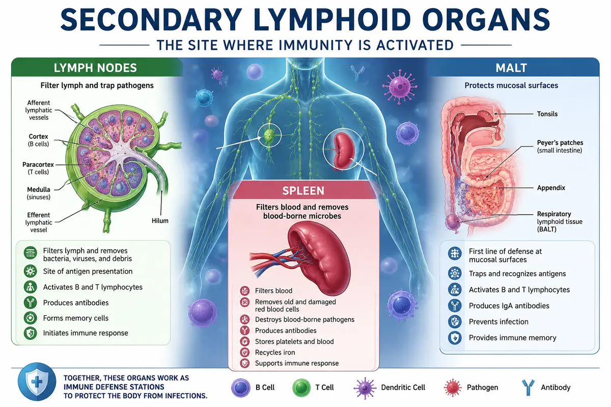
Similarly, lymph nodes, spleen, and mucosa-associated lymphoid tissues (MALT) are places where immune cells gather, detect harmful microbes, and coordinate an immune response to protect the body.
Secondary lymphoid organs are specialized organs where mature lymphocytes (B cells and T cells) encounter foreign antigens, become activated, multiply, and initiate the body’s immune response.
Main Secondary Lymphoid Organs: Lymph Nodes – Filter lymph and trap pathogens. Spleen – Filters blood and removes blood-borne microbes; Mucosa-Associated Lymphoid Tissue (MALT) – Protects mucosal surfaces such as the digestive, respiratory, and urinary tracts.
Secondary Lymphoid Organs: The Site of Immune Response Initiation
The basic lymphoid system, which includes T lymphocytes in the thymus and B cells, monocytes, dendritic cells, and granulocytes in the bone marrow, is where lymphocytes and myeloid cells mature. However, in the microenvironments of secondary lymphoid organs (SLOs), they come into contact with antigen and trigger an immunological response.
The organs where mature B and T cells encounter foreign antigens (germs), become activated, proliferate, and initiate the immune response are known as secondary lymphoid organs. Unlike primary lymphoid organs, they do not produce or mature lymphocytes but provide a place for immune cells to interact with pathogens.
Secondary lymphoid organs are specialized organs where mature lymphocytes encounter antigens, become activated, proliferate, and initiate immune responses. The main secondary lymphoid organs are
- Lymph Nodes—Filter lymph and trap pathogens.
- Spleen – Filters blood and removes blood-borne microbes.
- Mucosa-Associated Lymphoid Tissue (MALT) – Protects mucosal surfaces such as the digestive, respiratory, and urinary tracts.
Lymph Node
Lymph nodes are the most specialized SLOs. Lymph nodes are entirely dedicated to controlling an immune response, in contrast to the spleen, which also controls the flow and destiny of red blood cells. They are bean-shaped, enclosed structures made up of networks of stromal cells that are densely populated with dendritic, macrophage, and lymphocyte cells.
The first organized lymphoid structure to come into contact with antigens that enter the tissue voids is the lymph node, which is connected to both blood and lymphatic vessels. The lymph node offers the perfect microenvironment for antigen-lymphocyte interactions as well as effective, well-organized cellular and humoral immune responses.
Structure of lymph node
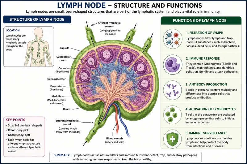
A lymph node is surrounded by a connective tissue capsule and is divided into three main regions:
1. Capsule
- Outermost tough connective tissue covering.
- Protects the lymph node.
- Sends inward projections called trabeculae.
2. Cortex (Outer Region)
- Located beneath the capsule.
- Contains lymphoid follicles rich in B lymphocytes.
- Some follicles contain germinal centers, where activated B cells multiply and differentiate into plasma cells and memory B cells.
3. Paracortex (Deep Cortex)
- Lies between the cortex and medulla.
- Rich in T lymphocytes and dendritic cells.
- Major site for antigen presentation and T-cell activation.
- Contains high endothelial venules (HEVs) through which lymphocytes enter from the blood.
4. Medulla (Inner Region)
- Central part of the lymph node. Contains medullary cords with plasma cells, B cells, T cells, and macrophages.
- Contains medullary sinuses, where lymph is filtered before leaving the node.
5. Lymphatic Vessels
- Afferent lymphatic vessels: Carry lymph into the lymph node.
- Efferent lymphatic vessel: Carries filtered lymph out through the hilum.
6. Hilum
- Indented region of the lymph node.
- Exit site for the efferent lymphatic vessel, blood vessels, and nerves.
Working of lymph node
The arriving (afferent) lymphatic vessels, which puncture a lymph node’s capsule multiple times and empty lymph into the subcapsular sinus, carry antigen from infected tissue to the lymph node’s cortex. On the surface of moving antigen-presenting cells, it either enters in particulate form or is processed and displayed as peptides.
Particulate antigen can be transferred to other antigen-presenting cells, such as B lymphocytes, by resident antigen-presenting cells in the subcapsular sinus or cortex. As an alternative, resident dendritic cells in the T-cell-rich paracortex may digest and present particulate antigen as peptide-MHC complexes on their cell surfaces.
T Cells in the Lymph Node
It takes 16 to 24 hours for each naïve T lymphocyte to explore every MHC-peptide combination that the antigen-presenting cells in a single lymph node present. The specialized endothelial cells of high endothelial venules (HEV), so named because they are bordered with unusually tall endothelial cells that give them a thicker appearance, allow naïve lymphocytes to penetrate the cortex of the lymph node.
After entering the lymph node, naïve T lymphocytes examine MHC-peptide antigen complexes on the paracortex’s dendritic cell surfaces. Fibroblast reticular cells (FRCs), a network of processes derived from stromal cells, traverse the paracortex. Through related adhesion molecules and chemokines, this network—known as the fibroblast reticular cell conduit system (FRCC)—directs T-cell motility.
Additionally, it appears that antigen-presenting cells encircle the conduits, providing circulating T cells with plenty of chance to explore their surfaces while being directed down the network. The likelihood that T cells will encounter their particular MHC-peptide combination is substantially increased by the existence of this specialized network.
T cells that peruse the lymph node but do not bind MHC-peptide combinations leave through the lymph node’s medulla’s efferent lymphatics rather than the blood. T cells will cease migrating and settle in the lymph node for a few days if their TCRs do attach to an MHC-peptide complex on an antigen-presenting cell they come across.
Here, it will multiply, and, in response to signals from the antigen-presenting cell itself, its offspring will develop into effector cells with a range of roles, giving CD8+ T cells the capacity to eliminate target cells. CD4 T cells have the ability to differentiate into a variety of effector cells, such as those that can further activate B cells, CD8+ T cells, and macrophages.
B Cells in the Lymph Node
Additionally, B cells are stimulated and develop into high-affinity plasma cells that secrete antibodies in the lymph node. Both direct interaction with an activated CD4 TH cell and antigen engagement by the B-cell receptor (BCR) are necessary for B cell activation. The lymph node’s structure facilitates both processes. Similar to T cells, B cells enter the lymph nodes through the HEV and travel through the blood and lymph on a daily basis.
They react to certain signals and chemokines that attract them to the lymph node follicle rather than the paracortex. They eventually rely on follicular dendritic cells (FDCs) for guidance, even though they may initially use the FRCC. FDCs play a crucial role in “presenting” antigen to developing B lymphocytes and preserving the integrity of the follicular and germinal centers.
The ability of B cells’ receptors to recognize unbound antigen sets them apart from T cells. The follicle is usually where a B lymphocyte encounters its antigen. The B cell gets partially activated and absorbs and processes the antigen if its BCR binds to it. As previously stated, B cells are actually specialized antigen-presenting cells that show CD4+ TH cells processed peptide-MHC complexes on their surface.
According to recent research, B cells that have effectively engaged and processed antigen alter their migration patterns and migrate to the T-cell-rich paracortex, where they are more likely to come into contact with an activated CD4+TH cell that will identify the MHC-antigen complex they present. They stay in contact with this TH cell for several hours after successfully engaging it, during which time they become fully activated and receive signals that cause B cell proliferation.
Some activated B cells differentiate directly into an antibody-producing cell (plasma cell), but others re-enter the follicle to establish a germinal center. A follicle that develops a germinal center is sometimes referred to as a secondary follicle; a follicle without a germinal center is sometimes referred to as a primary follicle.
Germinal centers are remarkable substructures that facilitate the generation of B cells with increased receptor affinities. In the germinal center, an antigen-specific B cell clone will proliferate and undergo somatic hypermutation of the genes coding for their antigen receptors.
Those receptors that retain the ability to bind antigen with the highest affinity survive and differentiate into plasma cells that travel to the medulla of the lymph node. Some will stay and release antibodies into the bloodstream; others will exit through the efferent lymphatics and take up residence in the bone marrow, where they will continue to release antibodies into circulation.
While some will leave through the efferent lymphatics and settle in the bone marrow, where they will continue to release antibodies into circulation, others will remain and release antibodies into the bloodstream.
Within 4 to 7 days of the initial infection, B lymphocytes are first activated, and the germinal center is established; however, germinal centers are active for at least three weeks. Particularly in the initial days following illness, lymph nodes enlarge noticeably and perhaps painfully. Both the proliferation of antigen-specific T and B lymphocytes within the lobe and an increase in the number of lymphocytes encouraged to migrate into the node are responsible for this swelling.
The Generation of Memory T and B Cells in the Lymph Node
In addition to the proliferation and functional differentiation of antigen-specific lymphocytes, the interactions between TH cells and APCs and between activated TH cells and activated B cells also produce memory T and B cells.
Memory T and B cells can leave the lymph node and move to and across tissues that initially came into contact with the pathogen, or they can settle in secondary lymphoid tissues. Central memory cells are memory T cells that live in secondary lymphoid organs. They differ from effector memory T cells that move between tissues in terms of phenotype and functional potential.
Function of Lymph Node
A lymph node is a small, bean-shaped secondary lymphoid organ that filters lymph and protects the body from infections. It acts as a meeting place where immune cells recognize foreign antigens and start the immune response.
1. Filters Lymph—Removes bacteria, viruses, toxins, and foreign particles from the lymph. Prevents pathogens from spreading throughout the body.
2. Traps Antigens—Captures foreign antigens carried in the lymph. Antigens are presented to lymphocytes to trigger an immune response.
3. Activates B and T Lymphocytes—B lymphocytes produce antibodies. T lymphocytes destroy infected cells and help regulate immunity.
4. Produces Antibodies—Activated B cells differentiate into plasma cells. Plasma cells produce antibodies that neutralize pathogens.
5. Initiates Immune Response-Serves as the main site where immune cells recognize pathogens and coordinate the body’s adaptive immune response.
6. Forms Memory Cells—Produces memory B cells and memory T cells. These cells provide faster protection if the same pathogen enters the body again.
7. Removes Dead Cells and Debris- Macrophages engulf and digest dead cells, damaged tissues, and microorganisms by phagocytosis.
Spleen
The spleen organizes the immune response against bloodborne pathogens.
The spleen, situated high in the left side of the abdominal cavity, is a large, ovoid secondary lymphoid organ that plays a major role in mounting immune responses to antigens in the bloodstream. Whereas lymph nodes are specialized for encounters between lymphocytes and antigens drained from local tissues, the spleen specializes in filtering blood and trapping blood-borne antigens; thus, it is particularly important in the response to systemic infections. Unlike the lymph nodes, the spleen is not supplied by lymphatic vessels.
Instead, blood-borne antigens and lymphocytes are carried into the spleen through the splenic artery and out via the splenic vein. Experiments with radioactively labeled lymphocytes show that more recirculating lymphocytes pass daily through the spleen than through all the lymph nodes combined.
Structure of the Spleen
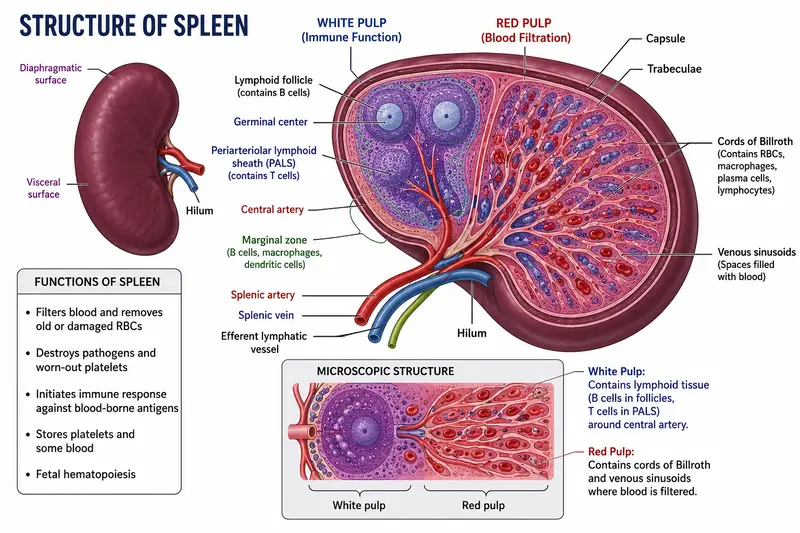
The spleen is the largest secondary lymphoid organ in the human body. It is located in the upper left side of the abdomen, just below the diaphragm and behind the stomach. Unlike lymph nodes, the spleen filters blood instead of lymph and plays an important role in immunity and the removal of old red blood cells. The spleen is enclosed by a connective tissue capsule and is divided into two major regions: white pulp and red pulp.
1. Capsule
- Thick connective tissue outer covering.
- Contains smooth muscle and elastic fibers.
- Protects the spleen.
- Gives rise to inward projections called trabeculae.
2. Trabeculae
- Connective tissue extensions from the capsule.
- Provide structural support.
- Carry blood vessels into the spleen.
3. White Pulp
- Appears white because it is rich in lymphocytes.
- Surrounds the central arteries.
- Composed mainly of the Periarteriolar Lymphoid Sheath (PALS)—rich in T lymphocytes—and lymphoid follicles—rich in B lymphocytes.
- Functions: Detects blood-borne antigens, activates B cells and T cells, and produces antibodies and memory cells.
4. Red Pulp
- Makes up about 75% of the spleen.
- Consists of splenic cords (cords of Billroth) and venous sinusoids.
- Function: Removes old or damaged red blood cells, destroys microorganisms, and stores platelets and blood. Macrophages recycle iron from aged red blood cells.
5. Hilum
- Indented region of the spleen.
- Entry and exit point for the splenic artery, splenic vein, lymphatic vessels, nerves
Working of Spleen
the initial line of protection against some bloodborne infections. The splenic artery allows blood-borne antigens and lymphocytes to enter the spleen, where they first interact with cells in the marginal zone. Dendritic cells in the marginal zone capture and process antigen before moving on to the PALS.
Additionally, local, specialized marginal zone B lymphocytes use complement receptors to bind antigen and deliver it to the follicles. Blood-borne B and T lymphocytes move to the follicles and PALS, respectively, after entering sinuses in the marginal zone.
Similar to what happens in the lymph node, the spleen’s adaptive immune response is triggered by similar events. In short, circulating naïve CD8+ and CD4+ T cells come into contact with antigen as MHC-peptide complexes on the surface of dendritic cells in the T-cell zone (PALS), while circulating naïve B cells come into contact with antigen in the follicles.
After being activated, CD4 TH cells aid B cells and CD8+ T cells that have also come into contact with antigen. Germinal centers are produced when certain TH cells and activated B cells return to follicles.
Whether a reticular network functions as significantly in the spleen as it does in the lymph node is unknown. However, it wouldn’t be shocking if a comparable conduit system existed, since T cells, dendritic cells, and B cells manage to effectively communicate within the spleen to start an immune response.
Animals can live comparatively healthy lives without a spleen, although losing one does have repercussions. Particularly in children, splenectomy (the surgical removal of a spleen) can result in overwhelming post-splenectomy infection (OPSI) syndrome, which is characterized by systemic bacterial infections (sepsis) primarily caused by Haemophilus influenzae, Neisseria meningitidis, and Streptococcus pneumoniae.
The importance the spleen plays in our immune response to pathogens that enter the circulation is highlighted by the fact that splenectomy can still result in an increased vulnerability to blood-borne bacterial infections, even though adults experience fewer side effects. It is crucial to understand that the removal of the spleen will also affect its other functions, such as hematopoiesis, thrombocyte storage, and iron metabolism.
Function of Spleen
The spleen is the largest secondary lymphoid organ. It filters blood instead of lymph and plays an essential role in immunity, blood filtration, and the removal of old red blood cells.
Functions of the Spleen
1. Filters Blood—Removes bacteria, viruses, parasites, and other foreign particles from the blood. Cleans the circulating blood.
2. Removes Old and Damaged Red Blood Cells—Macrophages destroy worn-out or damaged red blood cells. Helps maintain healthy blood circulation.
3. Initiates Immune Response—White pulp contains B lymphocytes and T lymphocytes. These cells recognize blood-borne antigens and start the body’s immune response.
4. Produces Antibodies—Activated B cells differentiate into plasma cells. Plasma cells produce antibodies that help destroy pathogens.
5. Stores Platelets and Blood—Acts as a reservoir for platelets and a small amount of blood. Stored blood and platelets can be released when needed.
6. Recycles Iron: Macrophages break down old red blood cells. Iron from hemoglobin is recycled and reused for the production of new red blood cells.
7. Hematopoiesis During Fetal Life—Before birth, the spleen helps produce blood cells. After birth, this function is mainly taken over by the bone marrow.
Mucosa-Associated Lymphoid Tissue (MALT)
When an antigen enters mucosal tissues, MALT coordinates the reaction.
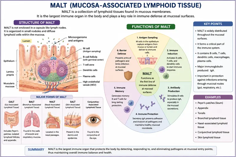
Secondary lymphoid microenvironments can form in organs other than the spleen and lymph nodes. Additionally, the epidermis and the mucosal membranes lining the respiratory, digestive, and urogenital systems contain T- and B-cell zones and lymphoid follicles.
The majority of infections enter through mucosal membranes, which have a combined surface area of over 400 m², or almost the size of a basketball court. Mucosa-associated lymphoid tissue (MALT) is a collection of organized lymphoid tissues that protect these delicate membrane surfaces.
Sometimes more precise names are given to lymphoid tissue associated with different mucosal areas. For example, the respiratory epithelium is called bronchus-associated lymphoid tissue (BALT) or nasal-associated lymphoid tissue (NALT), while the intestinal epithelium is called gut-associated lymphoid tissue (GALT).
The tonsils and adenoids (Waldeyer’s tonsil ring), the appendix, and Peyer’s patches, which are located within the intestinal lining and contain clearly defined follicles and T-cell zones, are examples of well-organized structures, whereas the structure of GALT is well described and ranges from loose, barely organized clusters of lymphoid cells in the lamina propria of intestinal villi.
The intestinal lining contains lymphoid cells in a number of different locations. T cells make up a large portion of the intraepithelial lymphocytes (IELs) found in the outer mucosal epithelial layer. Large numbers of B cells, plasma cells, activated T cells, and macrophages are seen in loose clusters in the lamina propria, which is located beneath the epithelial layer. A healthy child’s intestinal lamina propria has over 15,000 lymphoid follicles, according to microscopy.
Peyer’s patches are nodules made up of thirty to forty lymphoid follicles that protrude into the muscular layers immediately underneath the lamina propria. The lymphoid follicles that make up Peyer’s patches have the potential to grow into secondary follicles with germinal centers, just like those in other locations. Because MALT contains more antibody-producing plasma cells than the spleen, lymph nodes, and bone marrow combined, its overall functional significance in the body’s defense is undervalued.
Following their fusion with the pocket membrane, the vesicles transport antigens to the pocket’s clusters of lymphocytes and antigen-presenting cells, the most significant of which are dendritic cells. M cells carry antigen across the mucosal membrane, which ultimately triggers B cells to develop and release IgA. The body uses this class of antibody, which is abundant in secretions like milk, to fight off various infections at mucosal locations.
The Skin Is an Innate Immune Barrier and Also Includes Lymphoid Tissue
The largest organ in the body and a vital anatomical barrier against infections is the skin. Additionally, it is crucial for nonspecific (innate) defenses (Figure 2-13). Keratinocytes are specialized epithelial cells that make up the majority of the skin’s epidermal (outer) layer.
Numerous cytokines that may cause a local inflammatory response are secreted by these cells. Langerhans cells, skin-resident dendritic cells that absorb antigen by phagocytosis or endocytosis, are dispersed throughout the epithelial-cell matrix of the epidermis. After maturing, these Langerhans cells go from the epidermis to local lymph nodes, where they serve as powerful activators of naïve T lymphocytes.
In addition to Langerhans cells, the epidermis also contains intraepidermal lymphocytes, which are primarily T cells. According to some immunologists, these cells are best suited to fight infections that enter through the skin. There are also sporadic lymphocytes, dendritic cells, monocytes, macrophages, and possibly even hematopoietic stem cells in the skin’s underlying dermal layer.
The majority of skin lymphocytes seem to be either memory cells or previously activated cells, and many of them go to and from nearby draining lymph nodes that coordinate the reactions to pathogens that have penetrated the skin barrier.
Tertiary Lymphoid Tissues Also Organize and Maintain an Immune Response
Tertiary lymphoid tissue refers to the tissues that are the sites of infection. Antigen-activated lymphocytes in secondary lymphoid tissue have the ability to both reside as memory cells and return as effector cells to these organs (such as the brain, liver, and lung). Additionally, tertiary lymphoid tissues seem to be able to create specific microenvironments that arrange the returning lymphoid cells.
For example, researchers have discovered that the brain creates reticular systems that direct lymphocytes in response to a persistent infection with the protozoan that causes toxoplasmosis. Together, these findings demonstrate the immune system’s extraordinary plasticity and the close connection between anatomical structure and immunological function. The evolutionary links between immune systems and organs also demonstrate the conservation of structure/function relationships.
Lymphoid Organs Are Connected to Each Other and to Infected Tissue by Two Different Circulatory Systems: Blood and Lymphatics
The blood system and the lymphatic system are the two ways that immune cells move across tissues, making them the most mobile cells in the body. The blood is walled by endothelial cells that are very sensitive to inflammatory signals, and it has access to almost every organ and tissue.
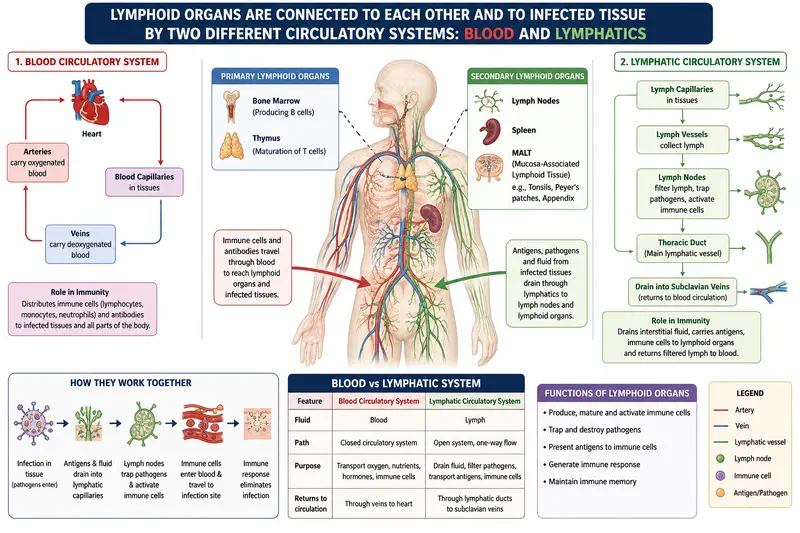
Within minutes, hemopoietic cells can move through the circulatory system, leaving the heart via active pumping networks (arteries) and returning to it via passive valve-based systems (veins). Through specialized blood arteries, the majority of lymphocytes enter secondary lymphoid organs and exit through the lymphatic system.
Immune cell trafficking, including the movement of antigens and antigen-presenting cells to secondary lymphoid organs and the departure of lymphocytes from lymph nodes, is significantly influenced by the lymphatic system, a network of thin-walled channels.
A protein-rich fluid called lymph, which is formed from the fluid component of blood called plasma, fills lymph veins and permeates the surrounding tissue through the thin capillary walls. An adult’s seepage can reach 2.9 liters or more over the course of a day, depending on their size and level of activity.
This fluid, known as interstitial fluid, bathes every cell and permeates every tissue. The tissue would enlarge and develop edema, which may potentially be fatal, if this fluid were not put back into circulation.
Because a large portion of the fluid is returned to the circulation through the venule walls, we do not suffer from such catastrophic edema. The delicate network of principal lymphatic capillaries receives the remaining interstitial fluid. One layer of loosely spaced endothelial cells makes up the walls of the principal vessels.
Fluids and even cells can enter the lymphatic network due to the major arteries’ permeable nature. The fluid, now known as lymph, passes from these capillaries into a sequence of progressively larger collecting vessels known as lymphatic vessels.
In the end, the lymphatic system returns all of its cells and fluid to the bloodstream. The thoracic duct, the largest lymphatic channel, flows into the left subclavian vein. With the exception of the right arm and right side of the head, it gathers lymph from every part of the body.
The right lymphatic duct, which empties into the right subclavian vein, receives lymph from these regions. The lymphatic system maintains steady-state fluid levels in the circulatory system by replenishing fluid lost from the blood.
Instead of the heart pumping lymph through the lymphatic system, the surrounding muscles move to provide a gradual, low-pressure lymph flow. As a result, exercise improves lymph circulation. Crucially, the lymphatic vessels have a number of one-way valves that guarantee lymph only flows in one direction.
When a foreign antigen enters the body, it is taken up by the lymphatic system, which eliminates all bodily fluids, and transported to different organized lymphoid tissues, including lymph nodes, where it is trapped. Lymph can also be accessed by antigen-presenting cells that take in and digest the antigen.
In actuality, lymph gradually gets enriched in particular leukocytes, such as lymphocytes, dendritic cells, and macrophages, as it travels from the tissues to lymphatic arteries. White blood cells and antigen are thereby transported by the lymphatic system from connective tissues to organized lymphoid tissues, where the lymphocytes can engage with the trapped antigen and become activated.
Chemokines are tiny chemicals that direct all immune cells as they move through tissues, blood, and lymph nodes. Stromal cells, antigen-presenting cells, lymphocytes, and granulocytes release these proteins, which create gradients that serve as attractants and guides for other immune cells that express a similarly varied range of chemokine receptors.
Immune cell movements can be highly precisely organized because of the interaction between particular chemokines and cells that express particular chemokine receptors. One extremely specialized secondary lymphoid organ is the lymph node.
Conclusion
Secondary lymphoid organs are the functional centers of the immune system, where mature B and T lymphocytes encounter foreign antigens and initiate adaptive immune responses. The lymph nodes filter lymph and activate immune cells, the spleen filters blood and removes blood-borne pathogens and aged red blood cells, and Mucosa-Associated Lymphoid Tissue (MALT) protects the body’s mucosal surfaces, which are the most common entry points for infectious microorganisms.
Together, these organs coordinate immune cell activation, antibody production, immune memory, and pathogen elimination, providing effective protection against infections while maintaining the body’s overall immune health. Understanding the structure and functions of secondary lymphoid organs is essential for appreciating how the immune system recognizes, responds to, and remembers harmful pathogens.
Frequently Asked University Questions (Previous 5 Years)
Long Answer Questions (8–15 Marks)
- Explain the structure of the lymph node with a neat, labeled diagram.
- Describe the histological structure and functions of the lymph node.
- Explain the organization of primary and secondary lymphoid organs.
- Discuss the structure and functions of secondary lymphoid organs.
- Explain the role of lymph nodes in adaptive immune response.
- Differentiate between primary and secondary lymphoid organs with suitable examples.
- Describe the flow of lymph through a lymph node with a diagram.
- Explain the distribution of B cells and T cells within a lymph node.
- Describe the immune functions of lymph nodes during infection.
- Explain the microanatomy of the lymph node and its clinical significance.
Short Notes (3–5 Marks)
- Structure of lymph node
- Cortex and medulla of lymph node
- Germinal center
- Paracortex
- Medullary cords
- Medullary sinuses
- Hilum of lymph node
- Afferent and efferent lymphatic vessels
- Functions of lymph node
- Secondary lymphoid organs
- Peyer’s patches
- MALT
- White pulp vs red pulp of the Spleen
- Lymphoid follicles
- Antigen presentation in lymph nodes.
Very Short Questions (1–2 Marks)
- Define a lymph node.
- What are secondary lymphoid organs?
- Name four secondary lymphoid organs.
- What is the function of a lymph node?
- Define germinal center.
- What is the hilum of a lymph node?
- What are afferent lymphatic vessels?
- What are efferent lymphatic vessels?
- Which cells are mainly present in the cortex?
- Which cells are mainly present in the paracortex?
- What is found in the medulla of a lymph node?
- Which lymphoid organ filters lymph?
- Which lymphoid organ filters blood?
- What is MALT?
- Name any two examples of MALT.
Most Important Questions
- Explain the structure of the lymph node with a labeled diagram.
- Differentiate between primary and secondary lymphoid organs.
- Explain the functions of the lymph node.
- Describe the pathway of lymph through a lymph node.
- Explain the role of B cells and T cells in the lymph node.
- Write a short note on secondary lymphoid organs.
Viva Questions
- Why are lymph nodes called “filters” of lymph?
- Why are there more afferent than efferent lymphatic vessels?
- Where are B lymphocytes located in a lymph node?
- Where are T lymphocytes located?
- What happens when an antigen enters a lymph node?
- Why do lymph nodes enlarge during infection?
- Which lymphoid organ filters blood instead of lymph?
- What is the importance of germinal centers?
FAQs
1. Is the spleen a secondary lymphoid organ?
Answer: Yes. The spleen is a secondary lymphoid organ.
It is the largest secondary lymphoid organ and plays a key role in the immune system by filtering blood, removing old or damaged red blood cells, and initiating immune responses against blood-borne pathogens. Unlike lymph nodes, the spleen filters blood rather than lymph.
2. What is a secondary lymphoid organ?
Answer: A secondary lymphoid organ is an organ where mature B and T lymphocytes encounter antigens, become activated, and initiate an immune response. These organs provide sites for immune cells to interact with pathogens and coordinate the body’s defense mechanisms.
Examples: Lymph nodes, spleen, and MALT (Mucosa-Associated Lymphoid Tissue).
3. Why is the spleen called a secondary lymphoid organ?
Answer: The spleen is called a secondary lymphoid organ because it is a site where mature B and T lymphocytes encounter antigens, become activated, and initiate immune responses. It filters blood, traps blood-borne pathogens, and helps produce antibodies, but it does not produce or mature lymphocytes (that occurs in primary lymphoid organs like the bone marrow and thymus).
4. Is the appendix a secondary lymphoid organ?
Answer: Yes. The appendix is considered a secondary lymphoid organ because it contains abundant lymphoid tissue (part of GALT—gut-associated lymphoid tissue) where mature lymphocytes encounter antigens and help initiate immune responses in the gastrointestinal tract.
5. Which organ is a secondary lymphoid organ?
Answer: Secondary lymphoid organs are the organs where mature lymphocytes encounter antigens and initiate immune responses.
Examples:
Lymph nodes
Spleen
MALT (Mucosa-Associated Lymphoid Tissue), including tonsils, Peyer’s patches, Appendix
References
- Kuby Immunology. Owen JA, Punt J, Stranford SA. Kuby Immunology. 9th ed. W.H. Freeman; 2019.
- Janeway’s Immunobiology. Murphy K, Weaver C. Janeway’s Immunobiology. 10th ed. Garland Science; 2022.
- Cellular and Molecular Immunology. Abul K. Abbas, Andrew H. Lichtman, Shiv Pillai. Cellular and Molecular Immunology. 11th ed. Elsevier; 2024.
- Roitt’s Essential Immunology. Peter J. Delves, Seamus J. Martin, Dennis R. Burton, and Ivan M. Roitt. Roitt’s Essential Immunology. 14th ed. Wiley-Blackwell; 2024.
- National Center for Biotechnology Information (NCBI Bookshelf). Immunobiology and Lymphoid Organs. NCBI Bookshelf.