
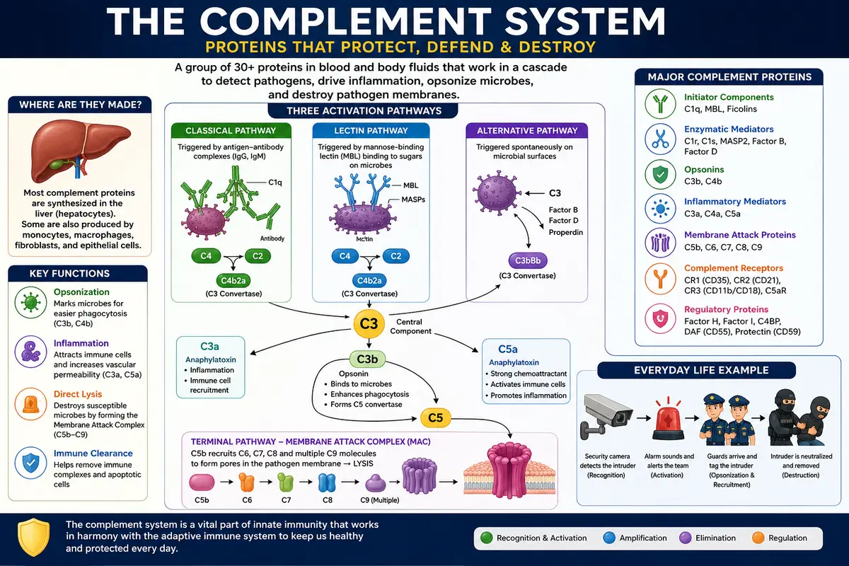
Learn about the which are the proteins involved in the complement system, their functions, activation pathways, and role in immunity with simple explanations and a day-to-day life example.
Introduction
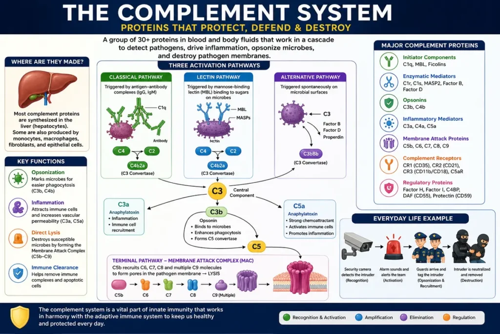
Think of your body like a city well protected. The city uses police officers, security cameras, alarms, and emergency teams to work together to find and take away threats. The human immune system also has a strong defense network called the complement system. It consists of a group of proteins, primarily in the blood and other body fluids, that work together to recognize and destroy harmful microorganisms.
The complement system consists of more than 30 proteins, such as complement components, regulatory proteins, and receptors. These proteins are normally present in an inactive form and are activated upon detection of pathogens such as bacteria, viruses, fungi, or other foreign substances. Once switched on, they act in an orderly sequence, often called a “cascade,” in which one protein activates another.
Suppose, for example, a thief enters a residential society. The security camera picks up the thief, the alarm sounds, security guards are called, and other emergency teams arrive to contain and remove the intruder. Complement proteins also recognize or react to microbes, recruit immune cells to the site of infection, enhance inflammation, opsonize microbes, making them easier for immune cells to engulf, and, in some cases, form the membrane attack complex (MAC), which may interfere with the membranes of certain microbes.
The major complement proteins are usually designated C1 thru C9, and other important components are Factor B, Factor D, Factor H, Factor I, properdin, and a number of regulatory proteins. These components are involved in three major activation pathways, the classical, lectin, and alternative pathways, which ultimately converge on the activation of C3, a central component of the complement system.
Therefore, knowledge of the proteins involved in the complement system is important, as each protein has a specific role in recognition, activation, inflammation, opsonization, and destruction of pathogens. Together they are an important part of the body’s innate immune defense and work closely with the adaptive immune system.
Which are the proteins involved in the complement system?
Researchers have since learned that complement’s activity results from interactions among a complex suite of more than 30 glycoproteins. Most of the complement components are synthesized in the liver by hepatocytes, although some are also synthesized by other cell types, including blood monocytes, tissue macrophages, fibroblasts, and epithelial cells of the gastrointestinal and genito-urinary tracts. Complement components constitute about 15% of the globulin protein fraction of plasma and may reach a total concentration of 3 mg/ml. In addition, several of the regulatory elements of the system are on cell membranes, so the term “complement” has come to include glycoproteins found in the blood plasma and on cell membranes.
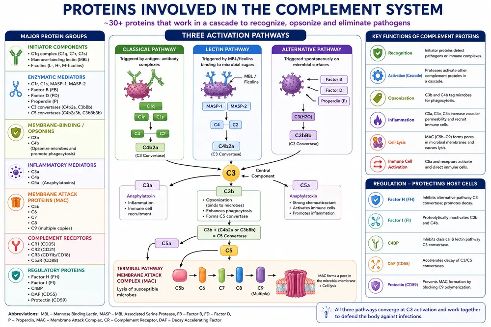
The components of complement are grouped into seven functional categories.
1. Initiator complement components
These proteins start their own complement cascades through binding to specific soluble or membrane-bound molecules. Binding to their activating ligand causes them to undergo a conformational change with a resulting change in biological activity. The initiator class of complement components includes the C1q complex, mannose-binding lectin (MBL), and the ficolins.
2. Enzymatic mediators
Some of the complement components (e.g., Clr, Cls, MASP2, and factor B) are proteolytic enzymes that can cleave and activate other members of the complement cascade. Some of these proteases are activated by binding to other macromolecules and changing conformation. Others are inactive until cleaved by another protease enzyme and are therefore called zymogens: proteins activated by proteolytic cleavage. The two enzyme complexes that cleave complement components C3 and C5, the C3 and C5 convertases, occupy places of central importance in the complement cascades.
3. Membrane-binding components or opsonins
When the complement cascade is triggered, a number of proteins are cleaved into two pieces, each of which has a specific function. C3b and C4b, the larger fragments of C3 and C4, respectively, also act as opsonins. By binding to microbial cells, they act as tags that facilitate binding to phagocytic cells that express receptors for C3b or C4b, thus promoting phagocytosis. The larger fragment of a cleaved complement component is generally designated with the suffix “b,” and the smaller with the suffix “a.” One exception to this rule is that the larger, enzymatically active form of the C2 component is designated as C2a.
4. Inflammatory mediators
Some small complement fragments are inflammatory mediators. These fragments bind to receptors on endothelial cells lining the small blood vessels and cause an increase in capillary diameter, thus improving the blood supply to the area in which they are released. They also recruit other cells to the site of tissue insult. “Against protection”—substances that cause anaphylaxis. These fragments are called “anaphylatoxins,” because such effects may be harmful in excess. For example, C3a, C5a, and C4a.
5. Membrane attack proteins.
Membrane attack complex (MAC) proteins insert themselves into the membranes of invading microorganisms and form pores in the membrane, leading to lysis of the pathogen. The complement components of the MAC are C5b, C6, C7, C8, and multiple copies of C9.
6. Complement receptor proteins.
Certain cell processes are signaled by receptor molecules on cell surfaces that bind complement proteins. For instance, certain complement receptors, like CR1, attach to complement components, like C3b, on the surface of pathogens, causing the C3-bound pathogen to be phagocytosed. Neutrophil degranulation and inflammation are triggered when the complement component C5a binds to C5aR receptors on neutrophils. “R” is used to name complement receptors, such as CR1, CR2, and C5aR.
7. Regulatory complement components.
Host cells are protected from unintended complement-mediated lysis by the presence of membrane-bound as well as soluble regulatory proteins. These regulatory proteins include Factor I, which degrades C3b, and Protectin, which inhibits the formation of the MAC on host cells.
The components of the complement system, their activation through three main channels, the effector roles of the complement cascade molecules, and their interactions with other cellular and molecular elements of innate and adaptive immunity. The evolution of the different complement proteins, the mechanisms that control the activity of these complement components, and the evasive tactics that infections have developed to evade complement destruction.
Conclusion
A crucial component of the body’s immune system, the complement system is made up of over thirty proteins that cooperate in a meticulously controlled cascade. The activation of C3, which is essential to complement-mediated immunity, is the final point of convergence for the three main activation pathways: the lectin pathway, the alternative pathway, and the classical pathway. Through opsonization, inflammation, immune cell recruitment, and direct destruction of microbial membranes, complement proteins aid the body in identifying and getting rid of pathogens.
Different complement proteins carry out specific tasks. While C3a and C5a encourage inflammation and draw immune cells to the infection site, C3b and C4b aid in marking bacteria for phagocytosis. The membrane assault complex (MAC), which is made up of the terminal components C5b, C6, C7, C8, and C9, has the ability to puncture the membranes of vulnerable microbes. Concurrently, regulatory proteins such as protectin (CD59), Factor I, and Factor H aid in preventing complement from harming healthy host cells.
Imagine a security force guarding a residential community to gain a basic understanding of the complement system. Certain proteins identify the intruder, some trigger an alarm and activate immune cells, others mark the intruder so security personnel can quickly recognize and eliminate it, and the MAC functions as an emergency team capable of causing direct harm to the intruder. Therefore, complement proteins collaborate as a coordinated defensive network rather than acting separately.
All things considered, the complement system serves as a crucial conduit between innate and adaptive immunity. It is crucial for efficient defense against infection because of its capacity to identify pathogens, boost immune responses, promote phagocytosis, and eliminate vulnerable microbes. In order to prevent needless inflammation and harm to the body’s own tissues, complement activity must be precisely regulated.
FAQs
1. What are complement proteins in the immune system?
Answer: Complement proteins are a group of more than 30 proteins in the blood and body fluids that help the immune system recognize and destroy harmful microorganisms.
They help in:
Opsonization—marking microbes for destruction.
Inflammation – attracting immune cells.
Pathogen destruction – forming the membrane attack complex (MAC).
2. How many proteins in the complement system?
Answer: The complement system consists of more than 30 proteins, including complement components, regulatory proteins, and complement receptors.
3. What are complement proteins?
Answer: Complement proteins are a group of more than 30 proteins found mainly in the blood that work together to recognize, attack, and eliminate harmful microorganisms. They also help promote inflammation and phagocytosis.
4. Which cells produce complement proteins?
Answer: Most complement proteins are produced by liver cells (hepatocytes). Some are also produced by monocytes, macrophages, fibroblasts, and epithelial cells.
5. What does complement protein C5a do?
Answer: C5a is a powerful inflammatory mediator and chemoattractant. It attracts immune cells such as neutrophils to the site of infection and helps promote inflammation.
References
- Abbas, A. K., Lichtman, A. H., & Pillai, S. (2023). Cellular and Molecular Immunology (10th ed.). Elsevier.
- Murphy, K., & Weaver, C. (2022). Janeway’s Immunobiology (10th ed.). W. W. Norton & Company.
- Punt, J., Stranford, S. A., Jones, P. P., & Owen, J. A. (2019). Kuby Immunology (8th ed.). W. H. Freeman.
- Kindt, T. J., Goldsby, R. A., & Osborne, B. A. (2007). Kuby Immunology (6th ed.). W. H. Freeman.
- Walport, M. J. (2001). Complement. New England Journal of Medicine, 344(14), 1058–1066.