
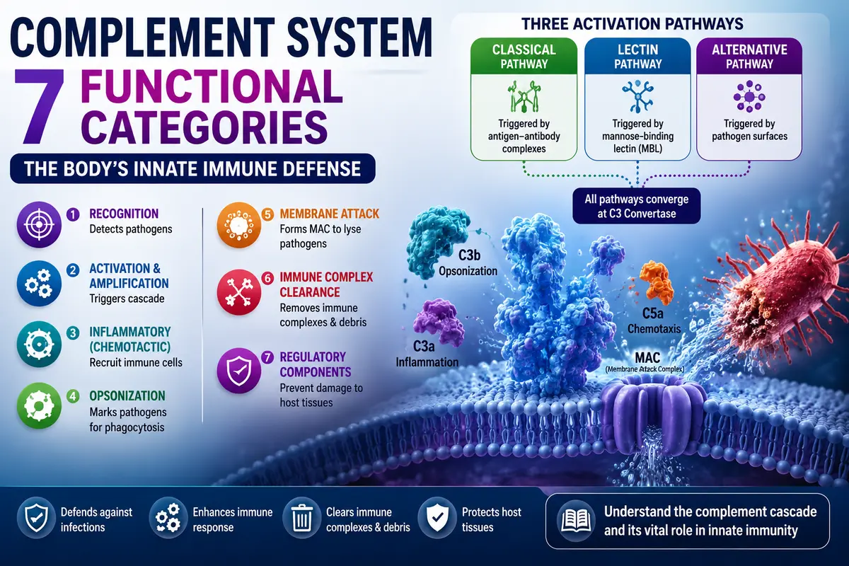
Learn about the seven functional categories of complement components, its components, functions, three activation pathways, seven functional categories, and role in innate immunity.
Introduction
Imagine a fire emergency response system. A smoke detector identifies the fire, an alarm alerts everyone, firefighters arrive to control it, the fire is extinguished, the area is cleaned, and safety officers ensure everything returns to normal. Each team has a specific role, but all work together to protect lives.
Similarly, the complement system is a group of more than 30 plasma proteins that work together as an important part of the innate immune system. These proteins remain inactive under normal conditions but become activated in a sequence called the complement cascade when pathogens enter the body. Each complement protein performs a specialized function to recognize microbes, enhance the immune response, recruit immune cells, destroy pathogens, remove immune complexes, and regulate the reaction to prevent damage to healthy tissues.
Based on their functions, complement components are classified into seven functional categories: recognition, activation and amplification, inflammatory (chemotactic), opsonization, membrane attack, immune complex clearance, and regulatory components. Together, these categories provide rapid and effective protection against infections while maintaining the body’s normal tissues.
Read our complete guide on immunology to understand the immune system from the basics.
Seven functional categories of complement components
A group of serum proteins known as complement (spelled with an e) works together with the innate and adaptive immune systems to eradicate blood and tissue infections. Complement proteins interact with each other in catalytic cascades, just as the elements of the blood clotting system.
Different components of complement bind and opsonize bacteria, making them vulnerable to receptor-mediated phagocytosis by macrophages that have membrane receptors for complement proteins. Additional complement proteins trigger inflammatory reactions, interact with adaptive immune system elements, remove immune complexes from the serum, and/or eradicate apoptotic cells.
Lastly, by making holes in microbial membranes, a Membrane Attack Complex (MAC) made of complement proteins directly kills some infections. The catastrophic effects of mutations in the genes encoding complement proteins and the variety of ways that microbes have developed to avoid complement highlight complement’s biological significance.
When Jules Bordet at the Institut Pasteur in Paris demonstrated that sheep antiserum to the bacterium Vibrio cholerae triggered lysis (membrane destruction) of the bacteria and that heating the antiserum reduced its bacteriolytic activity, complement research got underway in the 1890s.
Remarkably, adding fresh serum devoid of antibacterial antibodies restored the heated serum’s capacity to lyse the germs. Bordet deduced from this discovery that two distinct substances were needed for bacteriolysis: the heat-stable specific antibodies that attached to the bacterial surface and a second, heat-labile (sensitive) component that produced the lytic activity.
Bordet created red blood cell-specific antibodies in an attempt to purify this second, nonspecific component. He then utilized these antibodies in conjunction with purified serum fractions to determine which fractions worked with the antibodies to cause hemolysis, or the lysis of red blood cells. Similar studies were conducted separately in Berlin by renowned immunologist Paul Ehrlich, who defined complement as “the activity of blood serum that completes the action of antibody.”
Over the years, scientists have found that interactions between a complex set of over 30 glycoproteins are responsible for complement’s function. Hepatocytes create the majority of complement components in the liver, while other cell types such as blood monocytes, tissue macrophages, fibroblasts, and gastrointestinal and genitourinary tract epithelial cells also produce some.
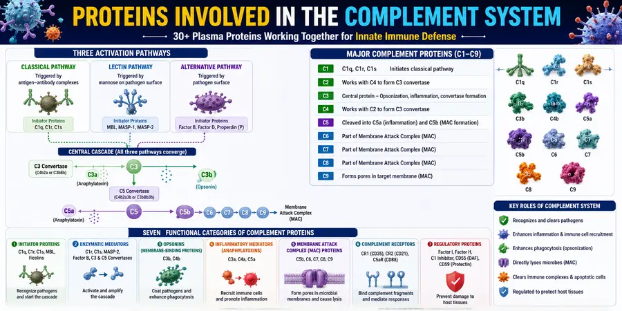
Complement components can have a total concentration of up to 3 mg/ml and make up around 15% of the globulin protein fraction in plasma. Furthermore, since a number of the system’s regulatory elements are found on cell membranes, glycoproteins found in both blood plasma and cell membranes are now included under the word complement. Seven functional categories can be used to categorize complement components.
1. Initiator complement components.
These proteins attach to specific soluble or membrane-bound compounds to start their corresponding complement cascades. Their biological activity changes as a result of conformational changes they experience after binding to their activating ligand. Initiator complement components include the ficolins, mannose-binding lectin (MBL), and the C1q complex.
2. Enzymatic mediators
Proteolytic enzymes that cleave and activate other elements of the complement cascade include Clr, Cls, MASP2, and factor B. By attaching to other macromolecules and changing their shape, some of these proteases are activated. Others, known as zymogens—proteins that are activated by proteolytic cleavage—remain dormant until they are broken down by another protease enzyme. The C3 and C5 convertases are the two enzyme complexes that cleave complement components C3 and C5, respectively. They play crucial roles in the complement cascades.
3. Membrane-binding components or opsonins.
When the complement cascade is activated, a number of proteins are split into two pieces, each of which subsequently assumes a specific function. The bigger pieces of C3 and C4, C3b and C4b, act as opsonins to increase phagocytosis by attaching to microbial cells and acting as binding tags for phagocytic cells that have C3b or C4b receptors. There is one exception to this rule: the bigger, enzymatically active form of the C2 component is called C2a. Generally speaking, the larger fragment of a cleaved complement component is identified with the suffix “b,” and the smaller with the suffix “a.”
4. Inflammatory mediators.
Certain tiny complement fragments function as mediators of inflammation. By attaching to receptors on endothelial cells lining the tiny blood capillaries and causing an increase in capillary width, these fragments improve the blood supply to the region in which they are released. Additionally, they draw additional cells to the tissue injury location. These particles are known as anaphylatoxins, which means “compounds that produce anaphylaxis (‘against protection’),” because such effects might be hazardous in excess. C3a, C5a, and C4a are a few examples.
5. Membrane attack proteins
The pathogen is lysed when the proteins of the membrane assault complex (MAC) penetrate the cell membranes of invasive microbes. The MAC’s complement components are several copies of C9, C6, C7, C8, and CSb.
6. Complement receptor proteins.
Certain cell processes are signaled by receptor molecules on cell surfaces that bind complement proteins. For instance, certain complement receptors, like CRI, attach to complement components, like C3b, on the surface of pathogens, causing the C3-bound pathogen to be phagocytosed. Neutrophil degranulation and inflammation are triggered when the complement component C5a binds to Call receptors on neutrophils. “R” is used to name complement receptors, such as CR1, CR2, and C5aR.
7. Regulatory complement components.
Both soluble and membrane-bound regulatory proteins protect host cells against unwanted complement-mediated lysis. Factor I, which breaks down C3b, and Protectin, which prevents the MAC from forming on host cells, are two examples of these regulatory proteins.
Conclusion
The complement system is a vital part of the innate immune system that works closely with the adaptive immune system to protect the body from infections. Although it consists of more than 30 plasma and membrane-associated proteins, each component has a specialized role in recognizing pathogens, amplifying immune responses, promoting phagocytosis, triggering inflammation, directly destroying microbes, clearing immune complexes, and regulating complement activity to prevent damage to healthy tissues.
The seven functional categories of complement components—initiator components, enzymatic mediators, membrane-binding components (opsonins), inflammatory mediators, membrane attack proteins, complement receptor proteins, and regulatory complement components—operate in a coordinated cascade to provide rapid and effective immune defense.
Together, they ensure that pathogens are detected, eliminated, and removed efficiently while maintaining immune balance and protecting host cells. Understanding these functional categories is essential for appreciating the complement system’s crucial role in host defense, inflammation, and immune regulation.
FAQs
1. What are complement components
Answer: Complement components are a group of more than 30 plasma and membrane-bound proteins that form the complement system, an important part of the innate immune system. They normally circulate in an inactive form and become activated in a cascade when pathogens invade the body. These proteins work together to recognize pathogens, promote inflammation, enhance phagocytosis (opsonization), destroy microbes through the Membrane Attack Complex (MAC), remove immune complexes, and regulate the immune response. The major complement components include C1 to C9, along with regulatory proteins and complement receptors.
2. What are the three types of complement?
Answer: The complement system is activated through three pathways:
Classical Pathway – Activated when antibodies (IgG or IgM) bind to antigens on a pathogen.
Lectin Pathway – Activated when mannose-binding lectin (MBL) binds to specific sugars on the surface of microorganisms.
Alternative Pathway – Activated directly by microbial surfaces without the need for antibodies.
All three pathways converge at the activation of C3 and ultimately lead to the formation of the Membrane Attack Complex (MAC), which destroys pathogens.
3. What are the main functions of the complement system?
Answer: The complement system recognizes pathogens, promotes inflammation, enhances phagocytosis (opsonization), destroys microbes through the Membrane Attack Complex (MAC), clears immune complexes, and regulates immune responses.
4. What is opsonization?
Answer: Opsonization is the process in which complement proteins, mainly C3b, coat pathogens to make them easier for phagocytic cells like macrophages and neutrophils to recognize and destroy.
5. Why is the complement system important?
Answer: The complement system provides a rapid first line of defense against infections, bridges innate and adaptive immunity, removes immune complexes, and helps maintain immune homeostasis.
References
- Abbas, A. K., Lichtman, A. H., & Pillai, S. (2024). Cellular and Molecular Immunology (11th ed.). Elsevier.
- Delves, P. J., Martin, S. J., Burton, D. R., & Roitt, I. M. (2023). Roitt’s Essential Immunology (14th ed.). Wiley-Blackwell.
- Murphy, K., & Weaver, C. (2022). Janeway’s Immunobiology (10th ed.). Garland Science, Taylor & Francis.
- Owen, J. A., Punt, J., & Stranford, S. A. (2021). Kuby Immunology (9th ed.). W. H. Freeman.
- Parham, P. (2021). The Immune System (5th ed.). Garland Science.
- Goldsby, R. A., Kindt, T. J., Osborne, B. A., & Kuby, J. (2003). Immunology (5th ed.). W. H. Freeman.
- NCBI Bookshelf – Immunobiology: The Complement System and Innate Immunity.
- Oxford Academic – The Complement System: An Introduction