
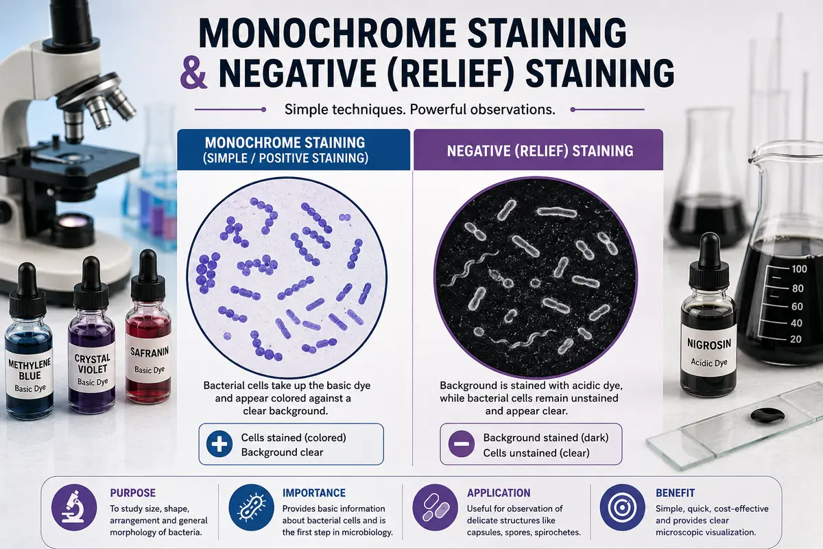
Learn about monochrome staining and negative (relief) staining techniques, their principles, dyes used, and importance in observing the size, shape, arrangement, and morphology of bacterial cells.
Introduction
Staining is not just a matter of adding color to microorganisms. It is a technique for making the invisible aspects of microbial life more visible and understandable. Monochrome and negative (relief) stains are among the simplest and at the same time the most informative methods of staining. These techniques make bacterial cells that are close to invisible visible and distinguishable under the microscope.
Monochrome staining involves using a single stain to give all bacterial cells the same color. This makes it easier to see the size, shape, arrangement, and general morphology of the cells. In contrast, negative or relief staining works differently: rather than staining the bacterial cell itself, it stains the background around the cell, leaving the cell itself relatively unstained. This creates a dramatic “relief” effect and permits the natural size and shape of the microorganism to be viewed with minimal distortion.
Because of their simplicity, clarity, and usefulness in the study of basic bacterial morphology, these staining techniques continue to be an important starting point in practical microbiology. They give students a clear visual introduction to the microscopic world and provide the basis for more sophisticated staining techniques.
Monochrome staining (Simple staining or positive staining)
“Simple staining” is the staining of bacteria by applying one solution of stain to a fixed smear. The fixed smear is flooded with a solution of a dye for a specified time. The solution is washed off with water, and the slide is blotted dry. The cells tend to stain evenly. Simple staining is also called “monochrome staining” because only one dye is used in a single application.
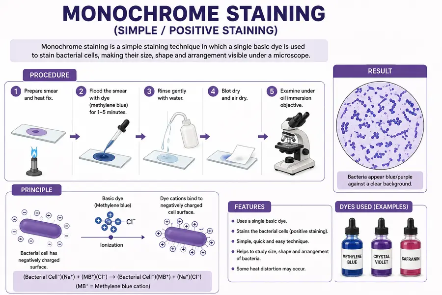
For this purpose, basic dyes like methylene blue, violet, crystal violet, safranine, malachite green, etc. are used. The reaction is based on the fact that the surface of the bacterial cell is overall acidic in character owing to the high concentration of carboxyl groups (-COOH) on the cell surface due to acidic amino acids. Therefore, when ionization of carboxy groups takes place, it imparts a net negative charge to the cell surface as per the following equation:
In nature, H gets swapped out for another positively charged ion, such as Na or K, and it forms a bond with oxygen to form water. Therefore, the staining process may involve an ion exchange response between the stain and active sites at the surface or inside the cell; for example, the dye’s colored ions may replace other ions on cellular components. Salt production with positively charged ions like Na or K may include specific chemical combinations of cell proteins or nucleic acids. As a result, we could see that the cell’s periphery contains both positively and negatively charged ions.
Methylene blue ionizes as follows during rehydration, producing both methylene blue and chloride ions. The coloring feature is possessed by the positively charged ions (MB). These ions take the place of K⁺ or Na⁺ on the cell surface. As a result, positively charged colored ions (MB) and negatively charged bacterial cell surfaces form ionic bonds, which stain the cell.
(Bacterial Cell–)(Na+) + (MB⁺)(Cl⁻)→(Bacterial Cell–)(MB⁺) + (Na⁺)(Cl⁻)
Procedure
(1) Prepare the smear and fix it with heat.
(2) Cover the film with methylene blue stain and allow it to react for 5 minutes.
(3) Wash the slide in running tap water, drain, blot, and air dry. Put a drop of cedarwood on the smear.
(4) Examine under the oil immersion objective lens.
Result: Organisms are stained blue in color.
Advantages of simple staining
Bacteria are difficult to see under microscope unless they are stained When organisms are stained with a particular stain, they take up the stain and can be seen more clearly when examined under a microscope. Staining of the organism helps in the study of morphological characteristics of the organism. Bacteria are seen as blue, violet, or red depending upon the stain used against the colorless background. The shape, size, and arrangements of cells of various bacteria can be studied. Thus, simple staining provides firsthand information regarding morphological characteristics of an unknown bacterium under examination.
Breed’s method for demonstration of bacteria in milk and McFadyean’s method for demonstration of anthrax bacilli are examples of practical applications.
Negative (Relief) staining
By using a process called negative staining, bacterial cells are left colorless while the backdrop is dyed with an acidic dye. Nigrosin is a common black dye. India Ink, Congored, Eosin, and other negative stains are examples. This method is applied to cells or structures that are challenging to directly stain. To show spirochaetes and capsules, negative staining is used.
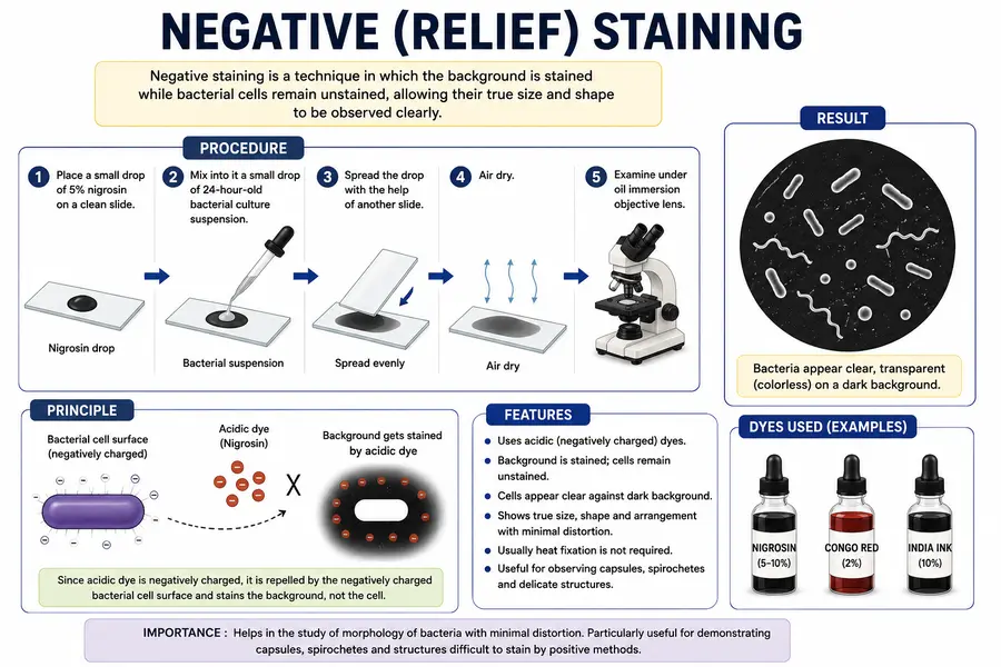
For the investigation of cell morphology, this method provides an advantage over direct or positive staining techniques. This is a result of the cells not being subjected to harsh physical or chemical treatments. Because acidic dyes, like cosin in sodium cosinate, have a negative charge, they do not mix with the negatively charged bacterial cell surface. However, it creates a deposit surrounding the cell, making the bacterial cell seem colorless against a dark background.
Procedure
(1) Place a small drop of 5% nigrosine on a slide
(2) Mix into it a small drop of a 24-hour-old bacterial cultural suspension.
(3) Spread the drop with the help of another slide.
(4) Air dry.
(5) Examine under an oil immersion objective lens.
Alternate procedure
(1) Prepare a smear and fix with heat.
(2) Place a drop of nigrosine at one edge of the slide.
(3) Spread by means of another slide over the bacterial film.
(4) Air dry.
(5) Examine under an oil immersion objective lens.
Result: Bacteria are seen as clear, transparent, colorless objects on a dark-black background. 5% nigrosin, 10% nigrosine, and 2% Congo red solutions are employed in negative staining. Phosphotungstic acid is the stain used to study the morphology of viruses in negative staining with virus particles. Electron dense phosphotungstate does not penetrate viruses.
Monochrome staining and Negative (Relief) staining difference
| Basis | Monochrome Staining | Negative (Relief) Staining |
|---|---|---|
| Definition | A staining technique in which bacterial cells are stained with a single dye. | A staining technique in which the background is stained while bacterial cells remain unstained. |
| Other names | Simple staining | Negative staining / Relief staining |
| Dye used | Basic dyes such as methylene blue, crystal violet, or safranin. | Acidic dyes such as nigrosin, India ink, or eosin. |
| Principle | Positively charged dye binds to the negatively charged bacterial cell surface. | Negatively charged dye is repelled by the negatively charged bacterial cell surface. |
| Appearance | Stained bacterial cells appear against a clear or lightly colored background. | Unstained bacterial cells appear as clear structures against a dark background. |
| Heat fixation | Generally required. | Usually not required. |
| Cell morphology | Shows shape, size, and arrangement of bacterial cells. | Shows the true size, shape, and arrangement with minimal distortion. |
| Cell distortion | Some distortion may occur due to heat fixation. | Very little distortion occurs. |
| Main purpose | To observe general bacterial morphology. | To observe accurate morphology and delicate structures such as capsules. |
| Example | Methylene blue staining | Nigrosin or India ink staining |
Conclusion
Monochrome staining and negative (relief) staining are simple and useful techniques for the microscopic study of bacteria. Monochrome staining helps visualize bacterial cells clearly by staining them with a single basic dye, whereas negative staining stains the background and leaves the bacterial cells unstained. Both techniques are valuable for studying the size, shape, arrangement, and general morphology of bacterial cells, with negative staining providing a more accurate representation with minimal cell distortion.
FAQs
1. What is monochrome staining?
Answer: Monochrome staining is a simple staining technique in which a single basic dye is used to stain bacterial cells, making their size, shape, and arrangement clearly visible under a microscope.
2. In monochrome staining, which stain can be used?
Answer: Stains used in monochrome staining:
Methylene blue
Crystal violet
Safranin
3. What is negative staining?
Answer: Negative staining is a staining technique in which the background is stained while the bacterial cells remain unstained, allowing their true size and shape to be observed clearly.
4. Why do we do negative staining?
Answer: Negative staining is done to observe the true size, shape, and arrangement of bacterial cells with minimal distortion.
5. What are positive and negative staining?
Answer: Positive staining: The bacterial cells are stained, while the background remains clear.
Negative staining: The background is stained, while the bacterial cells remain unstained.
References
- Cappuccino, J. G., & Welsh, C. T. (2017). Microbiology: A Laboratory Manual. Pearson.
- Leboffe, M. J., & Pierce, B. E. (2015). A Photographic Atlas for the Microbiology Laboratory. Morton Publishing Company.
- Harley, J. P. (2017). Laboratory Exercises in Microbiology. McGraw-Hill Education.
- Tortora, G. J., Funke, B. R., & Case, C. L. (2019). Microbiology: An Introduction. Pearson.
- Madigan, M. T., Bender, K. S., Buckley, D. H., Sattley, W. M., & Stahl, D. A. (2018). Brock Biology of Microorganisms. Pearson.