
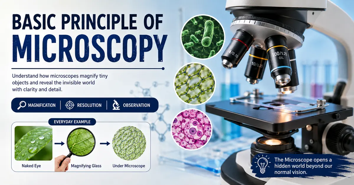
Learn what is the basic principle of microscopy is in simple language with easy day-to-day life examples. Perfect for students to understand magnification, resolution, and microscope working.
Introduction
Have you ever tried to read the tiny expiry date on a medicine strip or looked closely at the fine details of a flower using a magnifying glass? Suddenly, the small letters or tiny structures become much larger and easier to see. A microscope works on the same basic idea, but it is far more powerful than a simple magnifying glass.
Imagine placing a drop of pond water under a microscope. What looks like plain water to our naked eyes is actually filled with tiny living organisms swimming around. Without a microscope, these microorganisms remain completely invisible.
The basic principle of microscopy is to magnify very small objects and produce a clear, detailed image so they can be observed and studied. It uses light (or electrons in advanced microscopes) along with specially designed lenses to enlarge the specimen while improving its clarity. This allows scientists, doctors, and students to study cells, bacteria, fungi, blood cells, and many other microscopic structures that cannot be seen with the naked eye.
Microscopy is one of the most important techniques in biology, microbiology, medicine, and scientific research because it opens a hidden world that exists beyond our normal vision. In this article, you will learn the basic principle of microscopy in simple language with easy-to-understand examples from everyday life, making the concept easy to remember for both students and beginners.
Learn more about the history and science of microscopes.
History
Microorganisms are too small to be seen with the naked eye; thus, they can be seen with a microscope (micro = small, skopein = to see). In Van Leeuwenhoek’s day, looking through a microscope meant staring at the rainbow rings and shadows and many images. Modern microbiologists, nevertheless, have access to microscopes that create great-class magnifications from tens to thousands of times greater than those of Leeuwenhoek’s simple single lens.
In the seventh century, Anton van Leeuwenhoek utilized a rudimentary microscope with a single lens that resembled a magnifying glass. Around 1600, van Leeuwenhoek’s contemporaries, such as Robert Hooke, created compound microscopes with numerous lenses. But Joseph Jackson Lister, Joseph Lister’s father, created a much superior microscope. Lister’s microscope was improved upon in a number of ways, leading to the creation of the contemporary compound microscope that is currently utilized in microbiology labs.
The primary research instrument in microbiology, which studies microorganisms that are invisible to the unaided eye, is the microscope. A microscope may be defined as an optical instrument, consisting of a lens or combination of lenses, for making enlarged or magnified images of minute objects. Microscopes are of two categories depending upon the principle on which magnification is based: (1) light microscope or optical microscope and (II) electron microscope.
Certain basic characteristics of light and optics must be understood in order to comprehend microscopy. With this basis, many of the challenges faced in using a microscope will be more readily understood
Properties of light
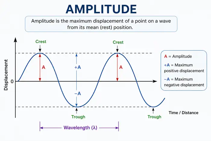
The speed of light is about 186,300 miles per second, or 3 × 10¹⁰ cm/second. According to wave theory, light is transported over space as waves of fluctuating electric and magnetic fields. Light waves are described in terms of their amplitude, frequency, and wavelength, as shown in the figure.
Explore the complete electromagnetic spectrum and visible light.
Amplitude
The largest displacement of a wave from equilibrium, represented by the crests and troughs of the resulting curves, is known as its amplitude.
A jump rope and a light wave can be compared. A posture of equilibrium is created when people pull on the rope and stretch it tightly. The highest displacement of the rope from the equilibrium position, as shown by the crests and troughs of the curves created, represents the wave’s amplitude when the rope is swung.
Frequency
This characteristic of a light wave is the quantity of vibrations that occur in a second. For instance, frequency is the number of times a trough or wave crosses a specific location in a second.
When two persons shake a rope, the pace at which the rope is shaken controls the frequency of the wave.
Wavelength and Frequency
The distance between two matching locations, such as the separation between two consecutive peaks or crests, is called a “wavelength.” Since the speed of light in a vacuum is constant and the frequency of light equals the number of wave crests or troughs that pass a place in a second, frequency and wavelength are inversely related.
Frequency = Velocity ÷ Wavelength
The wavelengths of light rays that make up the visible spectrum range, as shown in Figure , approximately 4,000 to 7,000 Å (400 to 700 nm).
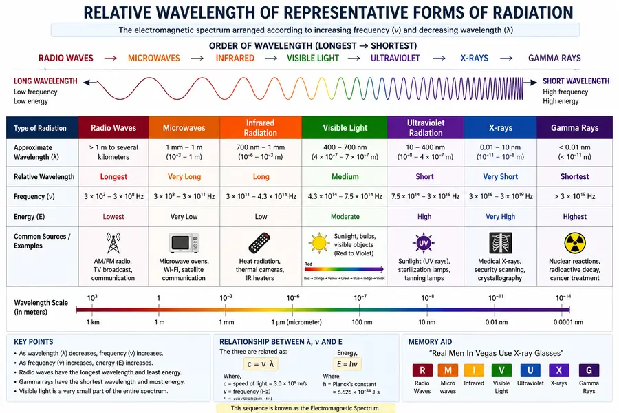
There are wavelengths that are either longer or shorter than the visible spectrum’s bounds. The wavelengths of ultraviolet light rays range from roughly 1,000 to 3,850 Å (100 to 385 nm). The wavelengths of infrared light are longer than those of visible light.
The resolving power (RP) of a microscope (its ability to expose the fine features of a specimen) relies on wavelength. Generally speaking, the resolving power increases with decreasing illuminating light wavelength. As a result, finer features are visible when illuminated by ultraviolet light as opposed to visible light.
Refractive Index and Refraction
When a beam of light crosses from one medium to another, refraction occurs, i.e., the ray is bent at contact (Figure 5.3). The refractive index controls the amount and direction of bending. The formula defines the refractive index, which is denoted by the Greek letter “n” (eta):
Refractive index (n) = Speed of light in a vacuum / Speed of light in medium being tested
Light is attenuated and bent toward normal when it travels from air into glass, a material with a larger “n.” As light leaves glass and returns to air, a medium with lower “n,” it accelerates and bends away from the normal. The index under regularly used materials.
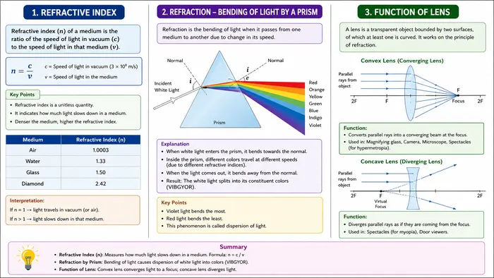
| Mounting Medium | Refractive Index (n) | Common Use |
|---|---|---|
| Water | 1.333 | Wet mounts, living specimens |
| Air | 1.00003 | – |
| Glass (an average) | 1.6 | – |
| Sandalwood oil | 1.51 | – |
| Crown oil | 1.55 | – |
| Paraffin oil | 1.47 | – |
| Olive oil | 1.47 | – |
| Glycerol (Glycerin) | 1.473 | Temporary mounts, fungi, algae |
| Glycerol Jelly | 1.47–1.48 | Mounting pollen, plant tissues |
| Canada Balsam | 1.52–1.53 | Permanent mounting of histological sections |
| DPX (Distrene Plasticizer Xylene) | 1.52 | Permanent mounting in light microscopy |
| Euparal | 1.48–1.49 | Permanent mounting of insects and plant specimens |
| Cedar Wood Oil | 1.515 | Oil immersion and special mounting applications |
| Immersion Oil | 1.515 | Oil immersion objective (100×) microscopy |
| Silicone Oil | 1.40–1.41 | Specialized microscopy applications |
| Hoyer’s Medium | 1.43–1.44 | Mounting fungi, mites, and small organisms |
Magnification
The material is magnified by the objective lens, which creates a true image (Figure 5.4). The real picture is transmitted through the microscope to the ocular lens, which magnifies the real image and produces an image seen by the observer, termed the virtual image.
The low-power lens, which has a magnification of 10X; the high-power lens, which has a magnification of 45X; and the oil immersion lens, which has a magnification of 100X, are the three objective lenses that are often mounted to a nosepiece in compound microscopes.
Learn how microscope objectives and magnification work in detail.
The magnification of the magnificatory ocular lens (which typically ranges from 5 to 20X) and the objective lens are multiplied to determine the overall magnification for the lens system. Thus, the utmost magnification produced by utilizing the oil immersion lens is approximately 2000X. The following magnifications are possible with a compound microscope:
Formula for Total Magnification
| Eyepiece (Ocular) | 2.5X Objective | 4X Objective | 10X Objective | 20X Objective | 45X Objective | 50X Objective | 100X Objective |
|---|---|---|---|---|---|---|---|
| 5X | 12.5X | 20X | 50X | 100X | 225X | 250X | 500X |
| 10X | 25X | 40X | 100X | 200X | 450X | 500X | 1000X |
| 15X | 37.5X | 60X | 150X | 300X | 675X | 750X | 1500X |
| 20X | 50X | 80X | 200X | 400X | 900X | 1000X | 2000X |
Conclusion
Microscopy has revolutionized the way we understand the invisible world. From observing bacteria and blood cells to studying plant tissues and microorganisms, the microscope has become an indispensable tool in biology, microbiology, medicine, and scientific research. Although the objects being studied are extremely small, the basic principle of microscopy is simple—it magnifies tiny specimens and produces a clear, detailed image by using light (or electrons) and a system of lenses.
Understanding fundamental concepts such as the properties of light, wavelength, frequency, refractive index, refraction, resolving power, and magnification makes it much easier to appreciate how a microscope works. Remember that a microscope is not just about making an object appear larger; it is equally important that the image is sharp and detailed. This is why resolution is often considered more important than magnification alone.
A simple day-to-day example is using a magnifying glass to read the fine print on a medicine strip or examining the intricate patterns on a butterfly’s wing. A microscope applies the same principle but with much greater precision, allowing us to explore a hidden world that is invisible to the naked eye.
Whether you are a school student, college learner, or an aspiring microbiologist, mastering the basic principle of microscopy provides a strong foundation for understanding advanced microscopic techniques such as bright-field microscopy, phase-contrast microscopy, fluorescence microscopy, confocal microscopy, and electron microscopy. Once you understand these basic principles, learning more complex concepts in microbiology and life sciences becomes much easier.
In short, microscopy is the gateway to the microscopic world, enabling scientists, doctors, and students to observe, analyze, and discover the fascinating structures and organisms that shape life around us. Understanding its principles not only strengthens your scientific knowledge but also helps you appreciate the remarkable technology that has transformed modern biology and medicine.
Explore advanced microscopy techniques and educational resources
Frequently Asked University Questions (Previous 5 Years)
Long Answer Questions (10–15 Marks)
- Explain the basic principle of microscopy with a neat, labeled diagram.
- Describe the construction, working principle, and applications of a compound microscope.
- Explain the properties of light and discuss their importance in microscopy.
- Describe wavelength, frequency, amplitude, and their relationship with suitable diagrams.
- Explain the principle of magnification and resolving power in microscopy.
- What is refractive index? Explain its significance in microscopy.
- Explain the principle and working of the oil immersion objective.
- Describe the factors affecting the resolving power of a microscope.
- Explain the formation of real and virtual images in a compound microscope.
- Differentiate between a light microscope and an electron microscope with suitable examples.
Short Answer Questions (3–5 Marks)
- Define microscopy.
- State the basic principle of microscopy.
- Define magnification and write its formula.
- What is resolving power?
- Define refractive index.
- Explain refraction of light.
- Write a note on wavelength.
- Define frequency.
- What is amplitude?
- State the relationship between wavelength and frequency.
- What is numerical aperture (NA)?
- List the objective lenses of a compound microscope.
- List the ocular lenses commonly used in microscopes.
- Write the formula for total magnification.
- Why is immersion oil used in microscopy?
Very Short Answer Questions (1–2 Marks)
- Define microscope.
- Expand the term NA.
- What is the SI unit of wavelength?
- Write the formula of frequency.
- State the speed of light.
- Write the formula for refractive index.
- Which objective lens requires immersion oil?
- Which objective provides the highest magnification?
- Which property of light determines resolving power?
- State the visible light wavelength range.
- Write the wavelength range of ultraviolet light.
- Name the scientist known as the Father of Microscopy.
- Name the scientist who developed the compound microscope.
- Define resolving power.
- What is the refractive index of air?
Numerical-Based Questions
- Calculate the total magnification when the objective lens is 45× and the eyepiece is 10×.
- A microscope has a 100× objective and 15× eyepiece. Find the total magnification.
- Calculate the frequency of light if its wavelength is given.
- Calculate the refractive index when the speed of light in a medium is provided.
Most Repeated University Questions
- Explain the basic principle of microscopy with a neat diagram.
- Define magnification and resolving power.
- Explain the properties of light in relation to microscopy.
- What is refractive index? Explain its importance in microscopy.
- Describe the construction and working of a compound microscope.
- Differentiate between magnification and resolution.
- Explain wavelength, frequency, and amplitude with suitable diagrams.
- What is the principle of the oil immersion objective?
- Derive the formula for total magnification of a compound microscope.
- Explain the factors affecting the resolving power of a microscope.
FAQs
1. What is the principle of magnification?
Answer: The principle of magnification is that a microscope uses objective and eyepiece lenses to enlarge the image of a small object, making it appear much larger than its actual size without changing the object itself.
Formula
2. What are the basic principles of light microscopy?
Answer: The basic principle of light microscopy is that visible light passes through or reflects from a specimen and is focused by objective and eyepiece lenses to produce a magnified, clear image. The quality of the image depends on magnification, resolution, contrast, and proper illumination.
3. What are the 4 magnifications of a microscope?
Answer: The four standard objective magnifications of a compound microscope are 4× (scanning), 10× (low power), 40× (high power), and 100× (oil immersion). These are used to observe specimens at progressively higher levels of detail.
4. What are 4x, 10x, 40x, and 100x magnifying lenses called?
Answer: 4× – Scanning Objective
10× – Low Power Objective
40× – High Power (High Dry) Objective
100× – Oil Immersion Objective
5. What are the different types of microscopy?
Answer: he two main types of microscopy are Light Microscopy and Electron Microscopy. Light microscopy uses visible light, while electron microscopy uses electrons to produce highly detailed images.
References
- Prescott’s Microbiology. Joanne M. Willey, Kathleen M. Sandman & Dorothy H. Wood. McGraw-Hill Education, 2020.
- Microbiology: A Laboratory Manual. James G. Cappuccino & Natalie Sherman. Pearson Education, 2020.
- Microbiology: Principles and Explorations. Jacquelyn G. Black. Wiley, 2018.
- Brock Biology of Microorganisms. Michael T. Madigan, Kelly S. Bender, Daniel H. Buckley, et al. Pearson, 2021.
- Ananthanarayan and Paniker’s Textbook of Microbiology. R. Ananthanarayan & C. K. Jayaram Paniker (Founding Authors). Universities Press, latest edition.
- Textbook of Microbiology. R. C. Dubey & D. K. Maheshwari. S. Chand Publishing.
- Olympus Life Science – Microscopy Resource Center Nikon Microscopy, Carl Zeiss Microscopy Knowledge Base, Encyclopaedia Britannica – Microscope