
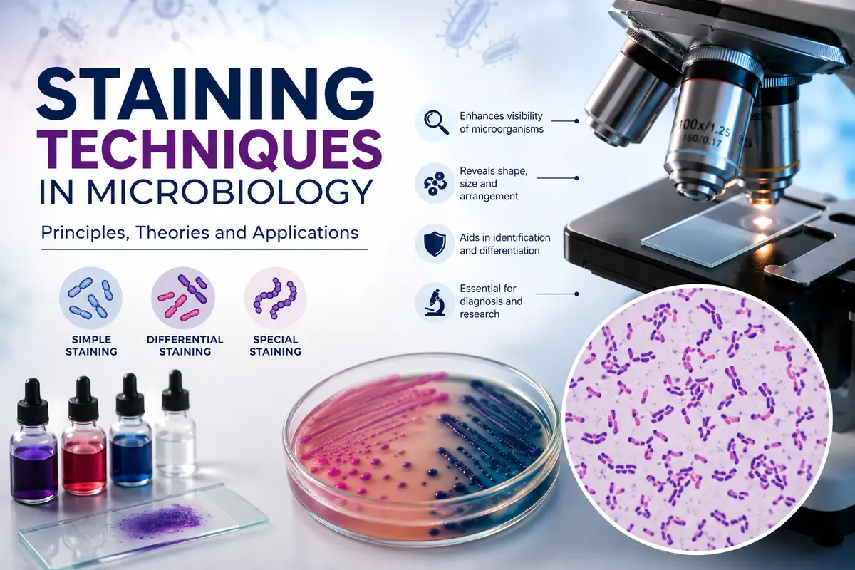
Master staining techniques in microbiology with easy explanations of the nature of staining processes, with physical and chemical theories, principles, applications, and exam-oriented notes.
Introduction
Staining is an essential technique in microbiology, histology, and pathology to view microscopic organisms and cells under a microscope. Most bacteria and other microorganisms are naturally transparent and almost colorless. They cannot be seen clearly without increasing the contrast between the specimen and the background. Staining is the use of special dyes that bind to the surface of the cell or to certain parts of the cell. This makes the organisms visible and enables scientists to study their shape, size, arrangement, and internal structures.
This is achieved by exploiting the interaction of the dye with the chemical composition of the cell. Certain stains can color the entire cell, whereas others have an affinity for particular structures such as the cell wall, nucleus, spores, or capsules. Depending on the objective, staining techniques may be simple, differential, or special and permit the identification and differentiation of a variety of microorganisms for research, diagnosis, and clinical applications.
One simple example of staining in everyday life is trying to see through a glass window with clear water writing on it, written transparently. The writing is almost invisible without any contrast. The hidden letters become clearly visible when the glass is sprayed with colored powder or paint. Bacteria are also naturally transparent. Stains work like the colored powder, so they can be seen under the microscope.
Another common example is adding a drop of food coloring to a glass of clear water. The color makes it easy to see the movement and distribution of the liquid. In the same way, stains help microbiologists to see microorganisms that would otherwise be invisible. Thus, it is an essential laboratory technique to increase visibility, improve contrast, aid in the identification of microorganisms, distinguish between different bacterial groups such as Gram-positive and Gram-negative bacteria, and disclose specific cellular structures, and it is important for disease diagnosis and microbiological research.
Principles of staining techniques in bacteriology
The protoplasmic matter of bacteria is quite definite. Therefore, bacteria are not easy to observe under a microscope unless they are stained. Bacterial protoplasm has nearly the same refractive index as the medium in which they grow, and therefore, they are not easily seen in the unstained state. Hence, staining techniques are very important in microbiology. The advantages of staining procedures are: (1) The cells are made more clearly visible after they are colored. (2) Differences between cells of different species and within the same species can be demonstrated by use of appropriate staining solutions, i.e., differential or selective staining.
Usually basic dyes are used for the purpose of staining in microbiology. The action of these stains can be enhanced by the use of intensifiers and mordants.
The nature of staining Processes (theories of Staining)
Many theories have been advanced to explain the phenomenon of staining. All attempt to explain the process on a purely physical or chemical basis.
(A) Physical theory of staining
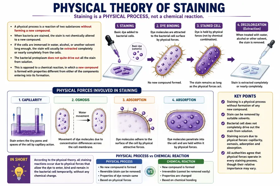
A physical process can be defined as a reaction of 2 substances without forming a new compound. When bacteria are stained, the stain is not shown to be chemically altered to a new compound. If the cells are immersed in water, alcohol, or another solvent long enough, the stain will usually be extracted completely or nearly completely from the cells. The bacterial protoplasm does not quite drive out all the stain from solution. This is generally supposed to be opposed to a chemical reaction, in which a new compound is formed with properties different from either of the components entering into its formation.
The supporters of the physical theory say that all the staining reactions can be explained on the basis of capillarity, osmosis, adsorption, and absorption. There appears to be no general agreement as to the amount of weight to be given to each force. All authorities are agreed on each occurrence of the staining process.
(B) Chemical theory of staining
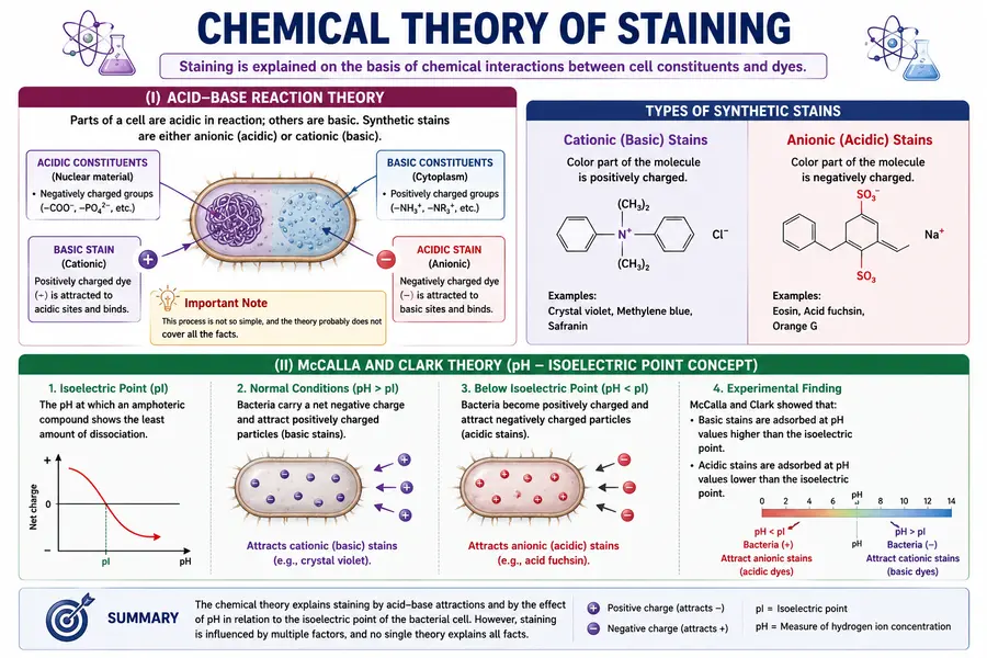
(I) Parts of a cell are acidic in reaction; others are basic. This fact led to the explanation of staining on a purely chemical basis. The synthetic stains are either anionic (acidic) or cationic (basic); i.e., the color part of the molecule is either the negative or the positive ion. The proponents of the theory said that the acidic constituents of the cell (nuclear material) reacted with basic stains and the basic constituents (cytoplasm) reacted with acidic stains. This process, however, is not so simple, and the theory probably does not cover all the facts.
(II) McCalla and Clark showed that basic stains were adsorbed at pH values higher than the isoelectric point and acidic stains at pH values lower than the isoelectric point (the isoelectric point is defined as that pH where an amphoteric compound shows the least amount of dissociation). Under normal conditions bacteria carry a negative electric charge and therefore attract positively charged particles. In the experiments of McCalla and Clark, bacteria in solutions with pH values below their isoelectric points are positively charged. Under these conditions they were able to absorb negatively charged stains such as acid fuchsin.
They concluded that the reaction of the stains with bacteria was an exchange process of adsorption attaining stoichiometrical proportions. On the basic side of the isoelectric point, basic stains served as cations to displace similarly charged ions from the bacterial system. On the acidic side of the isoelectric point, acidic stains served as anions to displace similarly charged ions from the bacterial system. The stains appeared to be reacting with the bacterial cell in the same positions as do inorganic cations and anions.
Applications of Staining Techniques in Microbiology
- Identification of Microorganisms—Staining helps identify bacteria, fungi, and other microorganisms based on their morphology and staining characteristics.
- Differentiation of Bacteria—Differential stains, such as the Gram stain and acid-fast stain, distinguish microorganisms into different groups for accurate identification.
- Study of Cell Morphology—Staining reveals the size, shape, arrangement, and structural details of microbial cells.
- Detection of Cellular Structures – Special staining techniques demonstrate bacterial structures such as spores, capsules, flagella, and inclusion bodies.
- Clinical Diagnosis of Infectious Diseases – Staining is widely used in medical laboratories to detect disease-causing microorganisms in clinical specimens.
- Microbiological Research—Researchers use staining techniques to study microbial physiology, taxonomy, and pathogenicity.
- Quality Control in Food and Water Testing—Staining assists in detecting microbial contamination in food products, beverages, and water samples.
- Pharmaceutical and Industrial Microbiology—Staining is used to monitor microbial contamination during the production of pharmaceuticals, vaccines, and other biological products.
- Teaching and Laboratory Education—Staining techniques are essential practical methods for demonstrating microbial characteristics to students in microbiology laboratories.
- Environmental Microbiology—Staining helps identify and study microorganisms present in soil, water, sewage, and other environmental samples.
Conclusion
Staining is a fundamental technique in microbiology that enhances the visibility of bacteria and other microorganisms by increasing the contrast between the specimen and the background. It helps in identifying the shape, size, arrangement, and specific cellular structures of microorganisms, making it indispensable for disease diagnosis, research, and laboratory investigations.
The staining process is explained by both physical and chemical theories, which describe how dyes interact with bacterial cells through mechanisms such as adsorption, absorption, ionic attraction, and pH-dependent reactions. Understanding the principles and theories of staining enables microbiologists to select appropriate staining methods for accurate observation, differentiation, and identification of microorganisms.
In summary, the evidence that is now available suggests that staining is most likely a combination of both physical and chemical processes.
FAQs
1. What are the principles of staining?
Answer: Increase Contrast: Staining makes transparent microorganisms visible by increasing the contrast between the cells and the background.
Affinity of Dyes: Stains bind to bacterial cells based on their chemical composition and electrical charge.
Basic and Acidic Dyes: Basic (cationic) dyes stain negatively charged bacterial cells, while acidic (anionic) dyes stain the background or positively charged cell components.
Role of Mordants: Mordants enhance stain binding and improve the intensity of staining.
Differentiation: Different staining methods help distinguish microorganisms based on their structural and chemical differences (e.g., Gram-positive and Gram-negative bacteria).
2. What is the difference between basic and acidic stains?
Answer: Basic stains color the bacterial cells, whereas acidic stains usually color the background.
3. What are the main types of staining?
Answer: The three main types are simple staining, differential staining, and special (structural) staining.
4. What is the chemical theory of staining?
Answer: The chemical theory explains that staining occurs because dyes chemically interact with acidic and basic components of the bacterial cell through ionic attraction.
5. What is the isoelectric point in staining?
Answer: The isoelectric point is the pH at which a cell has no net electrical charge, influencing how it interacts with acidic or basic dyes.
References
- Ananthanarayan and Paniker’s Textbook of Microbiology. (2023). Ananthanarayan and Paniker’s Textbook of Microbiology (11th ed.). Universities Press.
- Prescott’s Microbiology. (2020). Prescott’s Microbiology (11th ed.). McGraw-Hill Education.
- Brock Biology of Microorganisms. (2021). Brock Biology of Microorganisms (16th ed.). Pearson.
- Jawetz, Melnick & Adelberg’s Medical Microbiology (2023). Jawetz, Melnick & Adelberg’s Medical Microbiology (29th ed.). McGraw-Hill Education.
- Bailey & Scott’s Diagnostic Microbiology. (2019). Bailey & Scott’s Diagnostic Microbiology (15th ed.). Elsevier.
- Textbook of Microbiology. (2017). Textbook of Microbiology (10th ed.). Universities Press.
- Microbiology: A Laboratory Manual. (2020). Microbiology: A Laboratory Manual (12th ed.). Pearson.
- Microbiology. (2021). Microbiology: An Introduction (13th ed.). Pearson.