
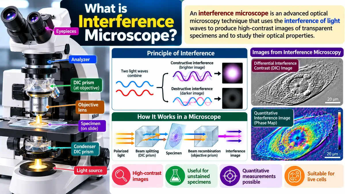
Learn what is interference microscopy?; its principle, working, types, applications, advantages, limitations, and differences from phase-contrast microscopy.

What Is Interference Microscopy?
Phase contrast microscopy relies on a single beam of light that passes through the specimen; interference microscopes have two beams of light that are combined after passing through the specimen to produce an interference pattern. An interference microscope can have higher numerical apertures, can vary the degree of contrast to suit the subject, and use colors to increase contrast. Interference microscopes can be used to observe living cells: phase changes that occur when light passes through the specimen are translated into changes in light intensity.
Principle of Interference Microscopy
The technique is based on the principle of interference of light. When two coherent light waves meet, they combine to produce either destructive interference of light.
- Constructive interference: Light waves reinforce each other, producing greater intensity.
- Destructive interference: Light waves partially or completely cancel each other, producing reduced intensity.
Differences in the optical path travelled by light through a specimen create measurable changes in the interference pattern.
The optical path length is approximately Optical Path Length = n X t.
Where:
n = refractive index of the medium or specimen
t = thickness of the specimen
Working of Interference Microscopy
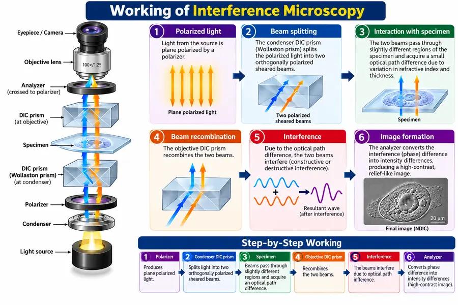
The working process involves the following steps:
- Light source: Light is directed towards the optical system.
- Beam splitting: In many interference microscopes, the light is divided into two beams.
- Specimen interaction: One beam interacts with the specimen, while the other acts as a reference beam.
- Beam recombination: The two beams are brought together.
- Interference formation: Differences in their optical paths produce an interference pattern.
- Image formation: The microscope’s optical system or camera records the resulting image for analysis.
Types of Interference Microscopy
1. Differential Interference Contrast (DIC) Microscopy
Produces high-contrast images with a relief-like appearance. It is useful for observing live cells, cell boundaries, and internal structures in transparent specimens.
Nomarski Differential Interference Contrast Microscopy (NDIC)

There are types of interference microscopes. NDIC is widely used by microbiologists. NDIC produces contrast images of unstained transparent specimens in three dimensions (3D). NDIC has a polarizing filter, an interference contrast condenser, and an analyzer plate. In NDIC, polarized light is split into two beams that travel at angles to each other and pass close together through the specimen.
The two beams of light are then combined and passed through the analyzer. When these two beams, which have been bent differently by the specimen, are recombined, these two beams produce interference. The pseudo‑3D image is produced because the two beams travel close together through the specimen, reducing depth effects.
Contrast in NDIC depends on the difference in how light bends across the specimen. Images seen through interference microscopes are normally brightly colored. Interference microscopes are useful for observation of unstained cells because interference microscopes produce images, with high contrast and striking topographic relief.
2. Quantitative Interference Microscopy
Measures optical phase differences to estimate specimen thickness, refractive index, or related properties when the appropriate optical and calibration information is available.
3. Multiple-Beam Interference Microscopy
Uses interference involving multiple reflected or transmitted beams for precise measurements of surfaces, thin films, and optical structures.
Applications of Interference Microscopy
| Sr. No. | Field | Applications |
|---|---|---|
| 1 | Microbiology | Studying transparent microorganisms and their cellular structures. |
| 2 | Cell Biology | Observing cell morphology, growth, movement, and division. |
| 3 | Medical Research | Examining biological cells and tissue specimens. |
| 4 | Materials Science | Measuring surface irregularities and microscopic material features. |
| 5 | Semiconductor Industry | Inspecting thin films, microstructures, and semiconductor surfaces. |
| 6 | Pharmaceutical Industry | Analyzing microscopic particles and the properties of drug formulations. |
| 7 | Biophysics | Measuring optical path differences and estimating cellular properties. |
| 8 | Cytology | Studying cell structure and changes in living cells. |
| 9 | Surface Analysis | Measuring surface profiles and small variations in height using suitable interferometric systems. |
| 10 | Thin-Film Analysis | Measuring the thickness and uniformity of thin films. |
| 11 | Quantitative Phase Imaging | Analyzing phase shifts caused by variations in specimen thickness and refractive index. |
| 12 | Research Laboratories | Investigating transparent specimens without staining in suitable microscopy methods. |
| 13 | Nanotechnology | Characterizing microscopic surface features and structures with appropriate interferometric instruments. |
| 14 | Industrial Quality Control | Detecting surface defects and evaluating the precision of manufactured components. |
| 15 | Live-Cell Imaging | Observing cellular changes over time while minimizing the need for staining. |
Advantages and Limitations of Interference Microscopy
| Sr. No. | Advantages | Limitations |
|---|---|---|
| 1 | Allows observation of transparent and unstained specimens. | Requires specialized optical components and equipment. |
| 2 | Produces high-contrast images of transparent biological specimens. | Optical alignment can be difficult. |
| 3 | Helps measure optical path differences accurately. | Measurements may be affected by vibrations and environmental changes. |
| 4 | Enables the study of living cells without staining in suitable methods. | Not all types of specimens produce clear interference images. |
| 5 | Helps estimate specimen thickness and refractive index using appropriate systems. | Quantitative measurements may require calibration and complex analysis. |
| 6 | Useful for studying cell morphology and movement. | Some methods produce shadowed or relief-like images that may be difficult to interpret. |
| 7 | Supports precise surface and thin-film measurements. | High-precision instruments can be expensive. |
| 8 | Useful in microbiology, medical research, and materials science. | Requires trained personnel for accurate operation and interpretation. |
| 9 | Can detect small variations in optical properties. | Results may be affected by specimen preparation and optical aberrations. |
| 10 | Provides valuable information for scientific research and industrial quality control. | Some applications require specialized software and additional analytical equipment. |
Difference Between Interference and Phase-Contrast Microscopy
| Sr. No. | Interference Microscopy | Phase-Contrast Microscopy |
|---|---|---|
| 1 | It is based on the interference of two or more light waves. | It is based on converting phase differences into intensity differences. |
| 2 | It uses separate light beams or wavefronts that are recombined. | It uses direct and diffracted light passing through a phase plate and related optical components. |
| 3 | It can produce interference fringes or contrast images. | It produces contrasting bright and dark regions in the specimen. |
| 4 | It can be used for quantitative measurements of optical path differences. | It is mainly used for qualitative observation of transparent specimens. |
| 5 | It can help estimate specimen thickness and refractive index with suitable calibration. | It is generally not used for direct quantitative thickness measurements. |
| 6 | Its optical arrangement may be relatively complex. | Its optical arrangement is generally simpler to use. |
| 7 | It may require precise alignment of the interfering beams. | It requires proper alignment of the condenser annulus and phase plate. |
| 8 | It is useful for studying optical properties, surface profiles, and transparent specimens. | It is especially useful for observing living cells and microorganisms. |
| 9 | Common types include differential interference contrast (DIC) and quantitative interferometric microscopy. | Commonly used as a standard phase-contrast light microscopy technique. |
| 10 | It is used in biology, materials science, surface analysis, and metrology. | It is widely used in microbiology, cell biology, and medical research. |
| 11 | Some methods produce a three-dimensional-like relief appearance, but this is not necessarily true topography. | Images may show halos around specimen boundaries. |
| 12 | It can provide precise optical measurements when the system is appropriately designed. | It is particularly effective for viewing unstained, transparent biological specimens. |
| Feature | Interference Microscopy | Phase-Contrast Microscopy |
|---|---|---|
| Basic principle | Interference between light waves | Conversion of phase differences into intensity differences |
| Image appearance | May show interference fringes or relief-like contrast | Often shows bright and dark cellular details |
| Quantitative measurement | Possible with suitable interferometric systems | Generally used for qualitative imaging |
| Common applications | Optical path, thickness, and surface measurements | Observation of living, transparent cells |
Conclusion
Interference microscopy is an important microscopy technique that converts differences in the optical paths of light into visible image contrast. It is widely useful in biological research, materials science, and precise optical measurements.
FAQs
1. What is interference microscopy?
Answer: Interference microscopy is an advanced optical microscopy technique that combines two light beams to produce interference patterns, enabling high-contrast imaging and the study of transparent, unstained specimens and their optical properties.
2. What is the principle of interference microscopy?
Answer: It works on the principle of light interference, in which two coherent light waves combine to produce constructive or destructive interference.
3.How does an interference microscope work?
Answer:It splits light into two beams, allows them to travel through different optical paths, and recombines them to form an interference pattern.
4. What are the main types of interference microscopy?
Answer: The main types include differential interference contrast (DIC) microscopy, quantitative interference microscopy, and multiple-beam interference microscopy.
5. Can interference microscopy observe living cells?
Answer: Yes, suitable interference microscopy techniques can observe living, unstained cells without requiring conventional staining.
References
- Wayne, R. O. (2019). Light and video microscopy (3rd ed.). Academic Press.
- Ruzin, S. E. (2024). Techniques in light microscopy. Oxford University Press
- Nolte, D. D. (2012). Optical interferometry for biology and medicine. Springer.
- Bradbury, S., & Evennett, P. J. (1996). Contrast techniques in light microscopy. BIOS Scientific Publishers. S. Bradbury a
- Murphy, D. B., & Davidson, M. W. (2012). Fundamentals of light microscopy and electronic imaging (2nd ed.). Wiley-Blackwell.