
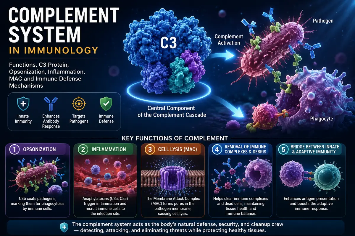
Explore the complement system in immunology, including What are functions of complement?, C3 protein, opsonization, inflammation, MAC, and immune defense mechanisms.
Introduction
An integral component of the body’s innate (natural) immune system is the complement system. It is made up of about thirty membrane-bound and plasma-bound proteins that are inactive and circulate in bodily fluids like blood. The complement cascade is a set of enzyme-mediated events that activate these proteins when pathogenic microbes, including bacteria, viruses, fungi, or parasites, enter the body.
Since these proteins support the functions of immune cells and antibodies, they increase the effectiveness of the body’s defense systems. The word “complement” implies boosting or aiding. The complement system not only targets pathogens directly but also increases inflammation, draws immune cells to the infection site, improves phagocytes’ capacity to engulf microbes, eliminates immune complexes and damaged cells, and acts as a crucial bridge between the innate and adaptive immune systems.
Comparing the complement system to a home security system is an easy way to comprehend it. Imagine having security cameras, an alarm system, security personnel, and a cleanup crew to keep your home safe. When an intruder tries to enter, the security cameras identify the threat, the alarm goes out right away, security personnel race to the scene and apprehend the invader, and then the cleanup crew cleans up any damage and reestablishes safety.
In the human body, the complement system serves a similar purpose. It detects invasive microorganisms, sends chemical signals that alert and attract white blood cells, marks pathogens for easy identification and destruction, directly kills some bacteria by creating holes in their cell membranes through the Membrane Attack Complex (MAC), and removes dead cells and immune complexes once the infection has been contained.
The complement system kills pathogens, removes damaged tissues, boosts the efficacy of antibodies, promotes inflammation when necessary, and aids in maintaining overall immunological balance as a result of these coordinated actions. The body is more susceptible to recurring infections and immune-related conditions when the complement system is not operating properly.
As a result, the complement system can be seen as the body’s natural defense, security, and cleanup crew. It constantly works to identify, combat, and eradicate dangerous microbes while preserving the integrity and health of bodily tissues.
What are functions of complement?
Complement has significant roles in innate immunity in addition to its well-known role in antibody-induced bacterial lysis. Many of these roles are mediated by soluble innate immune receptors like MBL and ficolins. C3 deletion animals exhibit greater susceptibility to bacterial and viral infections, demonstrating the critical role of C3b-mediated responses, including opsonization. Furthermore, recent studies have examined the functions of different complement components at the boundary between innate and adaptive immunity and discovered a number of ways that the release of active complement fragments influences the adaptive immune system.
Additionally, complement is crucial for the contraction phase of the adaptive immune response. Recent research has also indicated that complement is crucial for the removal of extra synapses during nervous system development. Below is a description of these different functions.
Complement Receptors Connect Complement-Tagged Pathogens to Effector Cells
The binding of complement fragments to cell surface complement receptors is essential for many of the complement system’s biological functions. Additionally, by facilitating the proteolysis of physiologically active complement components, certain complement receptors are crucial in controlling complement activity. Aspects of the innate and adaptive immune systems control the amounts of certain complement receptors. For instance, it has been demonstrated that activating phagocytic cells with different substances, such as the complement system’s anaphylatoxins, can increase the quantity of complement receptors by up to ten times.
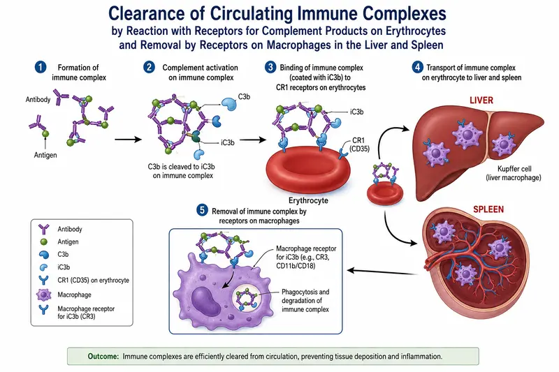
Therefore, we should familiarize ourselves with the receptors for complement components and their actions before delving into a description of the biological roles of complement. When a receptor has more than one name, we provide both in the initial introduction and then use the most popular name. Leukocytes and erythrocytes express CR1 (CD35), which has a strong affinity for C4b, C3b, and smaller C3b breakdown products. Immune complexes are bound by CR1 receptors on erythrocytes and transported to the liver, where phagocytes pick them up and eliminate them.
Proinflammatory chemicals including IL-1 and prostaglandins are secreted along with receptor-mediated phagocytosis when complement-opsonized microbial cells are bound by CR1 on phagocytes. CRI on B cells facilitates the uptake of C3b-bound antigen, which is then presented to T cells after being broken down in the B-cell lysosomal system. CR1 becomes involved in both the innate and adaptive immune responses as a result of this mechanism. By acting as an accelerator of the degradation of the C3 and C5 convertases and as a cofactor for the destructive cleavage of C3b and C4b on host cell membranes by factor I, CR1 also promotes the defense of host cells against the devastation of complement attack.
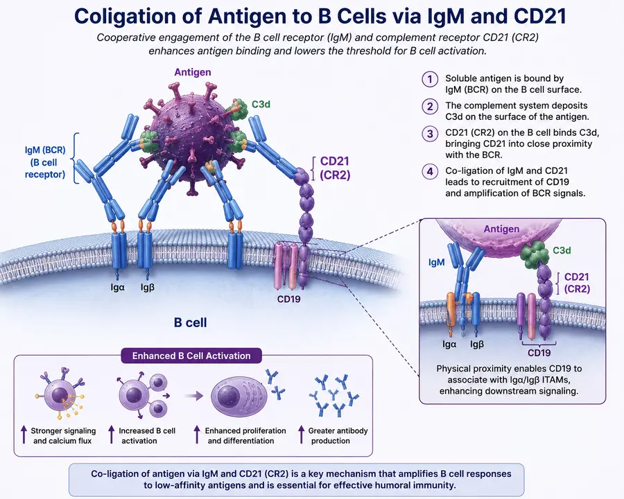
Endogenous proteases can degrade C3b when it is in solution or attached to the surface of cells. B cells express CD21 (CR2), which binds exclusively to iC3b, C3d, and C3dg, the breakdown products of C3b. The presence of CD21 on B cells allows the B cell to bind antigen via both the B-cell receptor and CD21 because C3b can establish covalent connections with antigens. The antigen concentration required for B-cell activation can be reduced by up to 100 times thanks to the capacity to concurrently co-engage antigen through two receptors. Patients with autoimmune conditions including systemic lupus erythematosus have been found to have deficiencies in CD21.
The phagocytosis of complement-coated antigens depends on CR3 (a complex of CD11b and CD18) and CR4 (a complex of CD11c and CD18). C3b and a number of its breakdown products, such as iC3b, C3c, and C3dg, are bound by CR3 and CR4. CRIg binds C3b as well. It is expressed by macrophages found in fixed tissues, such as the liver’s Kupffer cells. The discovery that CRIg-deficient mice are unable to effectively clear C3-opsonized particles highlights its significance in removing C3b-opsonized antigens from circulation in the liver. As a result, animals with this deficit are more likely to die from infections.
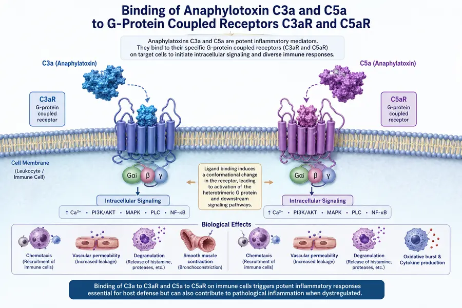
Members of the G protein coupled receptor (GPCR) family were initially identified in C3aR, C5aR, and C5L2. Following their binding to the tiny anaphylatoxins C3a and C5a, respectively, C3aR and C5aR mediate inflammatory activities. In addition to binding C5a, the C5L2 receptor shares structural similarities with C5aR and is expressed on some of the same cells. Nevertheless, C5L2 is not functionally linked to the G protein signaling pathway that C5aR uses; rather, signaling via C5L2 seems to regulate C5a signaling via C5aR, and animals lacking C5L2 exhibit increased inflammatory responses when C5a binds to its receptor.
More recently, it has been demonstrated that macrophages in the spleen’s marginal zone express SIGN-R1, a transmembrane lectin that can bind C1q. SIGN-R1 may bind carbohydrates found on the coat of the Gram-positive bacterium Staphylococcus pneumoniae and is found in aggregated form on the surface of macrophage cells. The bacterium finally becomes opsonized with C3b when SIGN-R1 attaches to bacterial polysaccharides, activating the C1q-binding capacity in the same or adjacent SIGN-R1 molecules.
Following their release from these macrophages, the opsonized bacteria are taken up by neighboring phagocytes, B cells, or dendritic cells. This peculiar method explains a long-standing issue with splenectomy patients: an increased vulnerability to S. pneumoniae infection.
Complement Enhances Host Defense
By creating the MAC, opsonizing potentially harmful microorganisms, and triggering an inflammatory response that aids in directing leukocytes to the infection site, complement proteins actively participate in host defense against infection.
MAC-Induced Cell Death
Complement’s role in causing cell death after the MAC is inserted into target cell membranes was the first function to be discussed. Erythrocytes were the target membranes in early MAC formation investigations, and huge holes comprising 17–19 molecules of C9 were found in this biological system. The creation of these pores in the cell membrane allowed ions and tiny molecules to flow freely. After a significant influx of water from the extracellular fluid, the cells lysed because the penetrated red blood cell membranes were unable to preserve osmotic integrity.
Subsequent research with nucleated eukaryotic cells, however, revealed that smaller pores may be created with just a few molecules of C9 and that in these situations, death happens by a form of apoptosis, or programmed cell death, once calcium enters the cytoplasm.
The idea that at least some MAC-targeted cells undergo apoptosis was further confirmed by the observation of nuclear fragmentation, a sign of apoptotic death, during MAC-induced lysis of nucleated cells. Apoptotic necrosis is the term used to describe MAC-induced apoptosis because more thorough investigations revealed that this type of apoptosis does not share all the molecular traits often associated with programmed cell death.
Since the membranes of eukaryotic cells contain a variety of components that work together to inactivate the complement pro-teins and shield the host cells from collateral damage during a complement-mediated attack on infectious microorganisms, killing eukaryotic cells with complement is actually quite challenging.
However, MACs can overpower the host’s defenses against MAC attack when complement component concentrations are high, and the resulting cell fragments can cause autoimmunity if they are present in high enough numbers. The complement system is thought to be a target for therapeutic intervention in autoimmune syndromes, and complement-mediated damage is a concern in a number of autoimmune illnesses.
Can a MAC attack be recovered from by a eukaryotic cell? MACs can be eliminated from the cell surface by either internalizing and breaking down the MAC-containing vesicles in intracellular lysosomes or by shedding MAC-containing membrane vesicles into the extracellular fluid, according to well-documented research. The cell can heal any damage to the membrane and restore its osmotic stability if the MAC is shed or absorbed soon enough after it first appears on the membrane. Tumor-specific antibody-directed complement-mediated lysis may be rendered ineffective by endocytosis or MAC shedding, which is an unpleasant consequence of this ability to rebound from MAC attack.
The degree to which certain bacteria are vulnerable to complement-induced lysis varies. Complement and antibodies are important components of the host’s defense against viruses, and they can be vital in preventing reinfection as well as limiting the spread of the virus during an acute infection. The majority of enveloped viruses are susceptible to complement-mediated lysis, including herpesviruses, paramyxoviruses like influenza, ortho-myxoviruses like those that cause measles and mumps, and retroviruses.
Because complement proteins are unable to pass through the bacterial cell wall and reach the membrane beyond, gram-positive bacteria effectively fend off complement attacks. On the other hand, complement attacks Gram-negative bacteria by first permeabilizing the outer layer and then lysing the inner bacterial membrane when the thin cell wall is destroyed.
In certain instances, it has been shown that the MAC localizes in areas where the inner and outer cell walls are next to one another and simultaneously breaches both. Patients who lack any of the complement components of the MAC are particularly susceptible to potentially deadly meningitis caused by the Gram-negative bacterium Neisseriammeningitidis, which is susceptible to MAC-induced lysis.
Promotion of Opsonization
The ability of antibodies and complement components (as well as other proteins) to wrap hazardous antigens that can subsequently be detected by Fc receptors (for antibodies) or complement receptors (for complement components) on phagocytic cells is known as opsonization. Complement receptor-mediated phagocytosis and antigen destruction occur when phagocytic cells bind complement-coated antigen.
Additionally, immunological complexes are bound by complement receptors on erythrocytes and subsequently transferred to the liver for macrophage phagocytosis. Opsonization may be the most physiologically significant role that complement components play, although it is less physically striking than MAC creation.
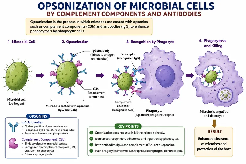
Complement and antibody opsonization offers vital defense against viral infection. By preventing the virus from attaching to receptors on the host cell, antibody and complement can form a thick protein coat that neutralizes viral infectivity. Additionally, it stimulates phagocytosis through complement receptors, which is followed by intracellular destruction of the broken-down particle.
Promotion of Inflammation
Thus far, we have concentrated on the functions of the major products of complement factor fragmentation: C5b in the development of the MAC and C3b and C4b in opsonization. Nonetheless, the smaller C3, C4, and C5 cleavage fragments—C3a, C4a, and C5a—are equally powerful and mediate crucial immune response events by functioning as anaphylatoxins or inflammatory inducers. Here, we shall pay particular attention to the actions of C3a and C5a.
Similar in structure, C3a and C5a are tiny proteins (about 9 kDa, or 74–77 amino acid residues in size) that both stimulate inflammation and act as chemoattractants for specific leukocyte types. On granulocytes, monocytes, macrophages, mast cells, endothelial cells, and some dendritic cells, C3a and C5a bind to G-protein-coupled activating receptors (C3aR for C3a and C5aR for C5a).
When these anaphylatoxins bind to their receptors on certain cells, a signaling cascade is set off that results in the release of soluble mediators, including TNF-a and IL-6. Leukocyte migration into the infection site is made possible by these cytokines’ localized increases in vascular permeability, which also cause a simultaneous increase in smooth muscle motility that aids in moving the released fluid to the site of injury.
A second round of inflammatory mediators, such as prostaglandins and histamines, are released as a result of these proinflammatory mediators’ promotion of phagocytosis of offending microorganisms and localized degranulation of granulocytes (neutrophils, basophils, and eosinophils). Inflammatory mediators accelerate lymphocyte migration into nearby lymph nodes, where the pathogen activates them. Fever and other systemic factors that reduce germ viability further reinforce this localized inflammatory response.
Clinicians looking for ways to reduce the pathological levels of inflammation experienced by patients with conditions like rheumatoid arthritis and systemic lupus erythematosus (SLE) have become interested in anaphylatoxins due to their central role in promoting physiologically significant inflammatory responses.
Complement Mediates the Interface Between Innate and Adaptive Immunities
Over the last 20 or 30 years, research has shown that the components of the complement system affect adaptive immunity through a variety of methods. Many of these discoveries are very recent, and research on the potential effects of complement component and regulatory protein binding on antigen-presenting cells, T cells, and B cells is still in its early stages.
Complement and Antigen-Presenting Cells
Every known complement receptor is expressed by macrophages, dendritic cells (DCs), and follicular dendritic cells (FDCs). MBL, C1q, C3b, and C4b can all bind to antigens and engage their corresponding receptors on antigen-presenting cells during the antigen recognition process. Signaling via these receptors then improves antigen absorption.
Furthermore, it has been demonstrated that antigen-presenting cells’ communication via the C5aR anaphylatoxin receptor modifies their motility and influences the generation of interleukins, particularly the cytokine IL-12. The T-cell response is typically skewed toward the Tal phenotype when an antigen-presenting cell produces IL-12. We must assume that numerous signaling pathways are being integrated to arrive at the final biological response to antigen and complement components because both induction and suppression of IL-12 production have been observed following activation of the anaphylatoxin receptors, depending on the route of antigen delivery, the nature of the antigen, and the maturation status of the antigen-presenting cell.
Complement and B-Cell-Mediated Humoral Immunity
Pepys demonstrated in 1972 that depriving mice of C3 reduced their T-dependent antigen-specific B-cell responses, suggesting that complement may be involved in the B-cell response’s beginning. It now seems that the complement receptor CD21 functioning as a coreceptor in antigen recognition was originally described in this groundbreaking finding.
Remember that the C3b molecule’s reactive thioester group interacts with hydroxyl or amino groups on the antigen’s surface to covalently “tag” the antigen. The immunoglobulin receptor is expressed on the B lymphocyte membrane along with the complement receptor CD21, the signaling protein CD19, and the transmembrane CD81 (tetraspanin, or TAPA molecule). CD21 is a receptor that is specific for both C3b and the products of C3b proteolytic breakdown, such as C3d (human) or C3Jg (mice). After C3b proteolysis, the C3d or C3dg fragments stay attached to the microbial surface if C3b was initially connected to it.
Complement and T-Cell-Mediated Immunity
Compared to B cells and antigen-presenting cells, the processes by which complement influences T-cell responses are less well understood. However, new research on mice treated with complement inhibitors or genetically defective in one or more complement components has produced some intriguing findings. For instance, CD4+ and CD8+ T cell responses are lower in animals lacking the C3 gene, suggesting that C3 signaling improves T-cell activity.
Additionally, after influenza infection, mice treated with C5aR signaling inhibitors generated fewer antigen-specific CD8+ T cells than wild-type mice, suggesting that C5a may function as a costimulator during CD8 T-cell activation, perhaps by boosting IL-12 production by antigen-presenting cells, as previously mentioned. These two investigations show that T-cell-mediated adaptive immunological responses may benefit from communication via complement components.
Although research on complement components’ regulation of T cells is still in its early stages, these intriguing results imply that there is still more to learn and that complement may have stronger effects on adaptive immunity than previously believed. The question of whether complement is directly affecting T cells or indirectly through impacts on antigen-presenting cells will need to be precisely characterized as these effects are investigated further.
Complement Aids in the Contraction Phase of the Immune Response
Only a small number of antigen-specific cells remain to provide immunological memory at the end of an adaptive immune response, as the majority of the lymphocytes produced during the initial proliferative phase undergo apoptosis (programmed cell death). Furthermore, the circulation and immunological organs may still contain soluble antigen-antibody complexes. Complement components are crucial in the proper disposal of these extra cells and immune complexes without causing additional inflammation if autoimmune illness is to be prevented.
Disposal of Apoptotic Cells and Bodies
On the outside of their plasma membranes, apoptotic cells express the phospholipid phosphatidyl serine. This phospholipid is typically limited to the cytoplasmic side of the membrane in healthy cells; this shift in position alerts the immune system that the cell is going through programmed cell death. Remarkably, studies have also shown that the surface membrane of apoptotic cells contains nucleic acids.
The production of outer membrane phosphatidylserine is rapidly followed by nuclear fragmentation and DNA cleavage. When apoptosis starts, the dying cell disintegrates into membrane-bound vesicles known as apop-totic bodies, which have surface-bound DNA and/or phosphatidyl serine on their external surfaces. Recent research has shown that C1q binds to DNA specifically.
Particularly, the complement component C1q binds apoptotic bodies and aids in their removal. C1q staining is limited to the apoptotic blebs, where DNA exposed on apoptotic membranes is particularly detected by C1q when light-sensitive keratinocytes (skin cells) are treated with UVB to start apoptosis and subsequently stained with anti-C1q antibodies. The conventional route is triggered by C1q deposition, and C3b subsequently opsonizes the apoptotic cells. Phagocytes then remove the apoptotic cells as a result.
When C1q is absent, the dying cells release the apoptotic membrane blebs as apoptotic bodies, which might subsequently function as antigens and trigger autoimmune reactions. As a result, compared to control mice, animals lacking C1q exhibit greater auto-antibody titers, higher mortality, and a higher incidence of glomerulonephritis, an autoimmune kidney disease. Immune complexes and a sizable number of apoptotic bodies are found in the kidneys of C1q mutant mice.
Disposal of Immune Complexes
As previously stated, the coating of soluble immune complexes with C3b makes it easier for CR1 on erythrocytes to bind to them. Since there are roughly 1,000,000 erythrocytes for every white blood cell, erythrocytes account for approximately 90% of the CR1 in blood, even though red blood cells express lower levels of CRI (100–1000 molecules per cell, depending on the age of the cell and the genetic constitution of the donor) than granulocytes (5 × 10¹ per cell).
Therefore, by transporting C3b-coated immune complexes to the liver and spleen, where they are separated from the red blood cells and phagocytosed, erythrocytes also contribute significantly to their removal. The discovery that patients with SLE, an autoimmune disease, have large amounts of immune complexes in their serum that are deposited in the tissues highlights the significance of the complement system in immune complex clearance. These tissue-deposited immune complexes activate complement, causing pathological inflammation in the afflicted tissues.
It may appear counterintuitive that there is a strong correlation between the incidence of SLE and C4 deficiency, as complement activation is linked to the pathogenesis of SLE. In fact, SLE develops in 90% of people who are totally deficient in C4. This paradox is resolved because deficiencies in the early components of complement result in lower levels of C3b deposited on the immune complexes. The subsequent inflammatory and cytolytic stages of complement activation can then be triggered as a result of this decrease, which also prevents their removal through C3b-mediated opsonization.
Complement Mediates CNS Synapse Elimination
It has also been demonstrated that complement has a significant role outside of the immune system. Growing neurons in the developing nervous system first create a comparatively large number of connections (synapses) with one another; as the nervous system develops, the number of those synapses is subsequently reduced.
Researchers studying the development of the mouse eye have shown that animals lacking C1q or C3 exhibit anatomical abnormalities in the visual nervous system and are unable to remove these early, excess synapses, suggesting that complement may be crucial in the process of synaptic remodeling.
Early in development, immature astrocytes—glial cells that support nervous system function and maintenance—provide signals that cause the production of complement components in mouse neurons to be up-regulated in healthy animals. Astrocyte function is therefore muted, and complement component expression is down-regulated as the animals age.
Nonetheless, it has been demonstrated that individuals with glaucoma exhibit abnormally elevated levels of complement components in the adult retina in the early stages of the condition, indicating that improper complement-mediated synaptic pruning could be a contributing cause. Additionally, C1q up-regulation has been seen in animal models of Alzheimer’s and ALS, which may indicate that several clinically significant neurodegenerative diseases are caused by improper complement activation.
Conclusion
An integral part of the innate immune system, the complement system supports adaptive immunity and offers quick and efficient defense against invasive infections. Opsonization of microorganisms, direct pathogen death via the Membrane Attack Complex (MAC), encouragement of inflammation, immune cell recruitment, removal of immune complexes and apoptotic cells, and immune response modulation are just a few of its many essential tasks.
Recent research has demonstrated its immunological function as well as its part in the development of the nervous system via synaptic pruning. Maintaining immunological homeostasis requires proper complement system control because both excessive activation and deficiency can result in inflammatory illnesses, autoimmune disorders, and recurring infections. Thus, the complement system ensures effective host defense, tissue protection, and general health by acting as a vital connection between innate and adaptive immunity.
FAQs
1. What are the functions of the complement group?
Answer: The main functions of the complement system are:
Opsonization—Coats pathogens to make them easier for phagocytes to recognize and destroy.
Cell Lysis—Destroys bacteria and other pathogens by forming the membrane attack complex (MAC).
Promotion of Inflammation – Releases inflammatory mediators (C3a and C5a) that increase blood vessel permeability and attract immune cells.
Chemotaxis – Recruits neutrophils and other white blood cells to the site of infection.
Clearance of Immune Complexes – Helps remove antigen–antibody complexes from the bloodstream.
Removal of Dead Cells – Aids in clearing apoptotic (dead) cells and damaged tissues.
Enhancement of Adaptive Immunity – Supports B-cell and T-cell responses, linking innate and adaptive immunity.
2. Which complement protein is used as an opsonin?
Answer: C3b – The principal complement opsonin that coats pathogens and enhances their phagocytosis by neutrophils and macrophages through complement receptors
3. Why is C3 a commonly measured complement protein?
Answer: C3 is commonly measured because it is the central and most abundant complement protein. It participates in all three complement activation pathways (classical, alternative, and lectin), so its level reflects the overall activity of the complement system. Decreased C3 levels may indicate complement activation, immune complex diseases, or recurrent infections.
4. What is the Membrane Attack Complex (MAC)?
Answer: The MAC is a complex of complement proteins that forms pores in the membranes of pathogens, leading to their destruction.
5. How does the complement system help remove immune complexes?
Answer: Complement proteins coat immune complexes, allowing their transport to the liver and spleen for removal by phagocytes.
References
- Owen, J. A., Punt, J., Stranford, S. A., & Jones, P. P. (2013). Kuby Immunology (7th ed.). W. H. Freeman and Company, New York.
- Abbas, A. K., Lichtman, A. H., & Pillai, S. (2024). Basic Immunology: Functions and Disorders of the Immune System (7th ed.). Elsevier.
- Murphy, K., & Weaver, C. (2022). Janeway’s Immunobiology (10th ed.). Garland Science, Taylor & Francis Group.
- Parham, P. (2021). The Immune System (5th ed.). W. W. Norton & Company.
- Delves, P. J., Martin, S. J., Burton, D. R., & Roitt, I. M. (2023). Roitt’s Essential Immunology (14th ed.). Wiley-Blackwell.
- Mescher, A. L. (2021). Junqueira’s Basic Histology: Text and Atlas (16th ed.). McGraw-Hill Education. (For immune cells and lymphoid tissues.)
- Tortora, G. J., Funke, B. R., & Case, C. L. (2024). Microbiology: An Introduction (14th ed.). Pearson.