
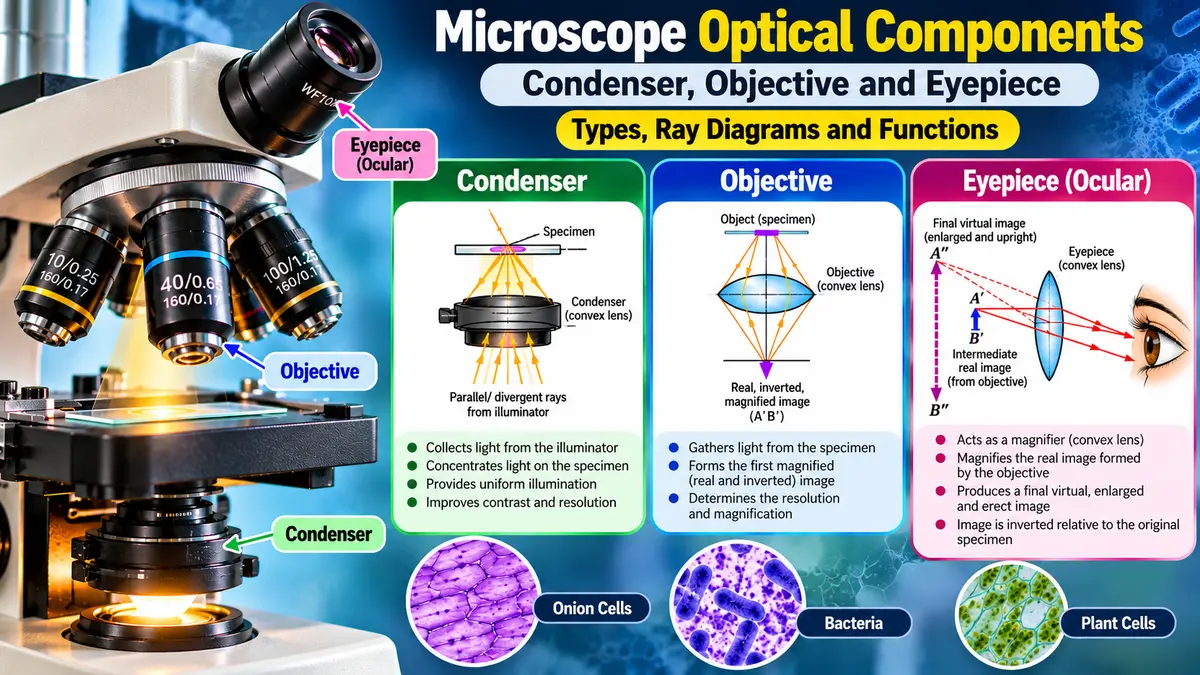
Explore the types, ray diagrams, and functions of microscope condensers, eyepieces, and objective lenses with simple, student-friendly explanations and examples.

Introduction
A Microscope Is More Than Just a Lens. A microscope does not just “make things bigger.” Every part inside it has a role. The condenser focuses light, the objective gathers light and creates the first magnified image, and the eyepiece makes that image bigger so our eyes can see it easily.
Have you ever used your mobile phone camera to zoom in on an object and seen how it becomes bigger and easier to make out?. Have you ever used a magnifying glass to read letters that are too small to see clearly? A microscope does something. It uses a setup of lenses to let us see things that are too small for our eyes to see properly. Think of a microscope like a camera system:
- Condenser—controls and directs the light
- Objective—captures and magnifies the specimen
- Eyepiece—magnifies the image for your eye.
Understanding these three components makes microscopy much easier.
In a compound microscope, light, lenses, and the specimen work together to produce a magnified image. Three important optical components play a role in this process: the condenser, objective, and eyepiece. Imagine them as a three-member team. The condenser is like a torch that focuses light where it is needed. The objective is like the camera lens that captures and magnifies the details of the specimen. Finally, the eyepiece is, like the zoom, that allows our eyes to see the enlarged image comfortably.
For example, when a student studying microbiology looks at bacteria or onion cells or a drop of water from a pond through a microscope, just putting the sample on the microscope is not enough. The condenser needs to give the amount of light. The objective has to make the sample bigger. The eyepiece has to make the picture even larger so we can see it clearly.
Condenser: The Light Manager of the Microscope
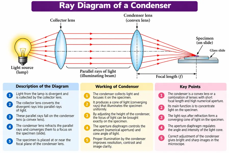
The condenser is a component that sits below the microscope stage. The condenser’s main job is to collect, concentrate, and focus light onto the specimen. Think of a flashlight shining on an object. If the flashlight’s light spreads over the object, it may look fuzzy. If the flashlight beam is aimed straight at the object, the details become sharp. That is what the condenser does.
Main Functions of a Condenser
- Collects light from the illumination system.
- Concentrates light on the specimen.
- Provides uniform illumination.
- Improves contrast and image clarity.
- Helps the objective lens receive appropriate light.
- Allows adjustment of the illumination cone using the iris diaphragm.
Basic Ray Diagram of a Condenser
Types of Condensers
Different condensers are designed for microscopy techniques. I think the Abbe condenser and the cardioid condenser are two types.
Abbe Condenser
The Abbe condenser is one of the commonly used condensers in ordinary bright-field microscopy. It was developed by the physicist Ernst Abbe, whose work played an important role, in the development of modern optical microscopy. An Abbe condenser generally consists of a system of lenses designed to concentrate light onto the specimen.
It is commonly used with bright-field microscopy. Routine laboratory microscopy, educational microscopes, biological specimen observation
The function of the Abbe condenser is to concentrate illumination on the specimen. The Abbe condenser produces a bright and relatively uniform field. It helps improve resolution and image contrast. It works effectively with routine bright-field microscopy. It allows control of illumination through the iris diaphragm.
Real-life Analogy: Suppose you are observing onion epidermal cells in a college practical. The light source provides illumination, then the Abbe condenser focuses that light on the onion peel, the objective collects the light coming from the specimen, and the eyepiece magnifies the resulting image. That entire process happens within seconds, but each optical component is performing a specific task.
Variable focus condenser
Variable‑focus condenser as a microscope condenser that lets me move the lens to focus the light on the specimen. This adjustment helps give the light, which makes the image clearer, brighter, and sharper. The condenser is placed below the microscope stage. By moving or adjusting the condenser, the position of the light beam can be changed. When the condenser is correctly focused, the light is concentrated precisely on the specimen, producing a brighter and sharper image.
Real-life Analogy: Suppose you are looking at bacteria in a stained smear using a high-power objective. If the condenser is not set up the way, the bacteria might look unclear. Changing the variable-focus condenser makes sure the light goes through the stained sample properly. This makes the bacterial cells easier to see.
Achromatic condenser
An achromatic condenser is a microscope condenser built to focus light onto a specimen while reducing aberration. An achromatic condenser uses a set of lenses made from kinds of glass so that light of all colors comes to almost the same focus. When white light passes through a lens, different colors can bend in different amounts. This can produce fringes around the specimen. An achromatic condenser corrects this problem by combining lenses with optical properties.
Real-life Analogy: When you photograph a black object against a bright sky using a poor-quality lens, you may sometimes see a purple or green-colored edge around the object. This is caused by chromatic aberration. A high-quality lens uses special optical elements to reduce this color error. Similarly, an achromatic condenser uses specially combined lenses to reduce color separation, allowing the microscope to illuminate the specimen more accurately.
Cardioid condenser
The cardioid condenser is a type of condenser that is mainly used in dark‑field microscopy. In a bright‑field setup, the cardioid condenser is different. The cardioid condenser is built so that direct light usually does not go into the objective. Instead, the cardioid condenser sends light at an angle toward the specimen. Why do we call it “cardioid”? The optical design of the cardioid condenser makes a pattern that looks like a heart shape. That shape is called a cardioid. The word “cardioid” comes from a word that means “heart shape.”
The functions of the cardioid condenser are to produce oblique illumination. It creates a dark background in dark-field microscopy. It makes small, thin, or low-contrast specimens appear bright. It also improves visibility of certain microorganisms. A cardioid condenser enhances the visibility of structures that may be difficult to see in ordinary bright-field microscopy.
Real-life Analogy: Imagine looking at a white dust particle on a background. The black background helps make the small particle much easier to see. Dark-field microscopy works in a way; the sample looks bright when placed against a dark background.
Eyepiece: The Window Through Which We See
The eyepiece, also known as the lens, is the lens system that the person looking into the microscope uses. It is found at the part of the microscope. The main job of this part is to make the image that the objective lens creates bigger. Imagine the objective as the person who takes a picture and the eyepiece as the person who makes that picture larger so you can look at it closely.
The curved upper part of the arm holds the body assembly to which one or more eyepieces (oculars) are attached. The oculars are used to magnify the image of the specimen produced by the objective and to correct certain distortions produced by the objective. Magnifying power of the ocular engraved on it. Common magnifications are 1X, 2X, 5X, 10X, 15X, and 20X. Microscopes with eyepieces for both eyes are called binocular microscopes. The body assembly has a series of mirrors and prisms.
Main Functions of Eyepiece
- Magnifies the intermediate image.
- Allows comfortable observation by the eye.
- Helps determine the final magnification.
- Provides a suitable field of view.
- Contributes to the overall optical performance of the microscope.
Types of Eyepieces
Different eyepiece designs have been developed to improve image quality and field of view.
| Eyepiece Type | General Feature |
|---|---|
| Huygens Eyepiece | Simple optical design |
| Hyperplane Eyepiece | Provide a clear and comfortable view of the specimen. |
| Compensating Eyepiece | Helps correct optical aberrations |
| Ramsden Eyepiece | Useful for basic optical systems |
| Kellner Eyepiece | Better correction and image quality |
| Orthoscopic Eyepiece | Good image flatness and low distortion |
| Wide-field Eyepiece | Provides a wider field of view |
Basic Ray Diagram of an Eyepiece
The objective first produces an intermediate image. The eyepiece then acts like a magnifying glass.
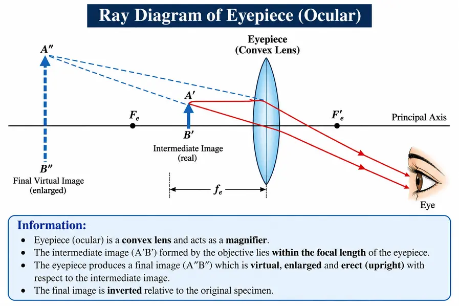
The eyepiece does not by itself create the picture of the sample. The objective lens makes the picture first, and the eyepiece makes that picture bigger. This is an important idea for students.
Objective Lens: The Main Magnifying Powerhouse
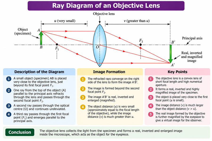
If the microscope were a team, the objective lens would be one of its important players. The objective is placed near the specimen. Is in charge of gathering light from the specimen and creating the first magnified image. Objective lenses often come with magnifications, giving the user the ability to look at the specimen from different levels.
The nose piece holds three to five objectives with lenses of differing magnifying power. It can be rotated to position any objective beneath the body assembly. The microscope must be parfocal, i.e., the image should remain in focus with the objective changing. The objective lens forms an enlarged real image with the microscope, and the ocular lens further magnifies to give a virtual image.
Most microscopes are equipped with three objectives with different magnifying powers: low power (10x), high power, or high-dry (45X), and oil immersion lens (100X). The objective lenses are used to gather (concentrate) the rays coming from the specimen and magnify this image.
Common Objective Magnifications:
| Objective | Magnification | Common Use |
|---|---|---|
| Scanning | 4× | Locating specimen |
| Low Power | 10× | General observation |
| High Power | 40× | Detailed cellular observation |
| Oil Immersion | 100× | Very high-resolution observation |
Main Functions of Objective Lens
The objective lens is one of the most important optical components of a microscope. It is located close to the specimen and performs the first stage of magnification.
- Collects light from the specimen – It gathers the light rays coming from the specimen.
- Forms the first image—The objective creates an enlarged real image of the specimen inside the microscope
- Magnifies the specimen—Different objectives provide different levels of magnification, such as 4×, 10×, 40×, and 100×.
- Reveals fine details—Higher-power objectives allow us to observe smaller structures, such as cells and microorganisms, in greater detail.
- Determines resolution—The objective plays a major role in the microscope’s resolution, or its ability to distinguish two closely spaced points.
- Works with the eyepiece—The objective first forms the magnified image, and the eyepiece further magnifies this image for the observer.
Types of Objective Lens
There are different types of objective lenses.
Achromatic Objective
The achromatic objective corrects chromatic aberrations and spherical aberrations. The achromatic objective is commonly used in educational microscopes and laboratory microscopes.
Plan Objective
Plan objectives are designed to provide a flat field of view, making them useful when the specimen needs to remain sharply focused across a larger area.
Fluorite/Semi-Apochromatic Objective
These provide improved optical correction and higher image quality compared with basic achromatic objectives.
Apochromatic Objective
Apochromatic objectives provide a high degree of correction for chromatic and spherical aberrations and are used when high-quality imaging is required.
Oil-Immersion Objective
The 100× oil-immersion objective is commonly used for very high magnification and resolution. A special immersion oil is placed between the coverslip and object.
Conclusion
A microscope is more than a tool that makes small things appear bigger. It is a built optical system where light is managed, focused, collected, and magnified in stages. The condenser gets the light ready. Sends it in the right direction. The objective lens creates the magnified image. Then the eyepiece takes that image. Makes it larger again for easy viewing.
Knowing what each part does helps explain how bright-field microscopy, dark-field microscopy, and high-resolution microscopy work. Each method uses the basic components but in different ways to show details in unique ways.
FAQs
1. What is the function of the condenser on a microscope?
Answer: The condenser collects and focuses light onto the specimen, providing proper and even illumination for a clear, bright image.
2. What does a condenser do in a microscope?
Answer: A condenser focuses light onto the specimen to provide clear and even illumination.
3. What does the eyepiece do on a microscope?
Answer: The eyepiece magnifies the image of the specimen so you can see it clearly.
4. What is the function of the eyepiece on a microscope?
Answer: The eyepiece magnifies the image formed by the objective lens and allows you to view the specimen.
5. What do the objectives do on a microscope?
Answer: The objective lenses magnify the specimen and form a detailed image for viewing.
References
- Microbiology: An Introduction — Tortora, G. J., Funke, B. R., & Case, C. L. Microbiology: An Introduction. Pearson.
- Prescott’s Microbiology — Willey, J. M., Sherwood, L., & Woolverton, C. J. Prescott’s Microbiology. McGraw-Hill.
- Benson’s Microbiological Applications — Brown, A. E. Benson’s Microbiological Applications: Laboratory Manual in General Microbiology. McGraw-Hill.
- Biological Science — Freeman, S. et al. Biological Science. Pearson.